

Role of NAD+ in regulating cellular and metabolic signaling pathways
Sara Amjad 1 11 , Sabah Nisar 2 11 , Ajaz A. Bhat 2, Ab Rauf Shah 3 , Michael P. Frenneaux 4, Khalid Fakhro 5 6, Mohammad Haris 2 7, Ravinder Reddy 8, Zoltan Patay 9, Joseph Baur 10,
Puneet Bagga 9
Molecular Metabolism, Volume 49, July 2021, 101195
Abstract
Background
Scope of review
Major conclusions
- NAD+ regulates energy metabolism, DNA damage repair, gene expression, and stress response.
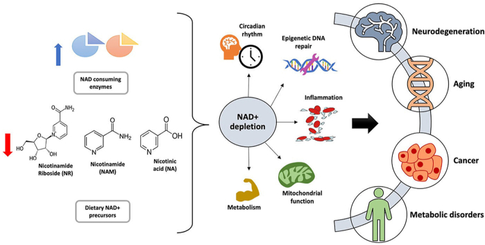
- NAD+ deterioration contributes to the progression of multiple metabolic disorders, cancers, and neurodegenerative diseases.
- Nicotinamide mononucleotide and nicotinamide riboside raise NAD+ levels in different tissues in preclinical models.
- Imaging studies on genetic models can illustrate the pathways of NAD+metabolism and their downstream functional effects.
- Human clinical trials to determine benefits of restoration of NAD+ by using NAD precursors are in progress.
2. NAD+ biosynthesis pathways

Figure 1. Representation of NAD+ biosynthesis pathways. Biosynthesis pathways and cellular metabolism of NAD+. NAD precursors such as NR, NA, NAM, and Trp provided by diet can be converted into NAD via three pathways. In the Preiss-Handler pathway, NA is converted into NAMN by NAPRT, NAMN is converted into NAAD by NAD by NMNATs, and NAAD is converted into NAD by NADSYN. In the de novo synthesis pathway, Trp is converted into QA in a series of steps, which is then converted into NAD by forming NAMN and NAAD. In contrast, in the salvage pathway, NR and NAM provided by diet are converted into NAD by forming NMN by enzymes NAMPT and NRK [2]. The equilibrium in each subcellular compartment such as the nucleus and mitochondria is determined by NAD/NADH redox ratios. ETC is a significant contributor to the conversion of NADH into NAD. Additionally, NAD-consuming enzymes such as PARPs and sirtuins catalyze NAM production in subcellular compartments, which can be used for NAD synthesis via the salvage pathway.
2.1. Preiss-Handler pathway
2.2. De novo biosynthesis pathway
2.3. Salvage pathway
3. Roles of NAD+ and sirtuins in cellular maintenance

Figure 2. Role of NAD in aging, neurodegeneration, and cancer. DNA damage caused by stress or aging activates PARP. PARP activation leads to reduced levels of cytosolic NAD and mitochondrial dysfunction, which contribute to aging and neurodegeneration. Any disturbance in levels of NAD+/NADH (redox homeostasis) can upregulate oncogenic signaling pathways, leading to tumorigenesis.
4. NAD+ in aging

Figure 3. NAD+ depletion with increasing age is caused by several factors such as inefficient metabolism or protein consumption, increased activity of NAD+-consuming enzymes CD38/PARP, mitochondrial dysfunction, DNA damage, cellular dysfunction, and NAMPT depletion. NAD+ can be restored by manipulating enzymes in NAD+ synthesis pathways. The NAD+ concentration can be replenished by increasing the activity of NAMPT, niacin, NMN, and NR. Moreover, in many cases, inhibition of PARP/CD38 also helps restore NAD+.
5. Role of NAD+ in neurodegenerative disorders

Figure 4. Altered NAD metabolism is associated with neurodegeneration. Axonal injury leads to the activation of SARM1, which reduces NAD + levels and leads to axonal degeneration. The overexpression of NMNAT1 inhibits SARM1 and protects injured axons. NAD levels are associated with axonal degradation, and impairment in the KYN pathway causes fluctuations in KYN pathway metabolite levels, which impairs the neurotransmission process and leads to neurodegeneration and the development of neurological disorders [201].
5.1. Alzheimer’s disease
5.2. Parkinson’s disease
5.3. Huntington’s disease
6. Role of NAD+ in metabolic disorders
6.1. Diabetes

Figure 5. The polyol pathway becomes highly active during hyperglycemia, leading to the massive production of NADH [202,203]. Consumption of NAD+ by PARPs results in NAD+ decline. During diabetes, accumulation of NADH and depletion of NAD+ leads to an increase in redox imbalance, which causes oxidative stress, decreased SIRT activity, decreased ATP production, and increased cell death.
6.2. Obesity
6.3. Hepatic steatosis and non-alcoholic fatty liver disease
6.4. Kidney diseases
7. NAD+ in cancer

Figure 6. NAD metabolism in cancer cells. Cancer cells rely on increased glycolysis rates for energy production and regenerate NAD+ by converting accumulating pyruvate into lactate to maintain glycolysis. Excess lactate accumulation in tumor cells increases the level of NADH relative to NAD and perturbs the NAD/NADH balance in cells [204]. In contrast, SIRT1 acts as an inactivator of HIFα and prevents its nuclear translocation. SIRT6 acts as a corepressor of HIFα to prevent the transcriptional process, and mitochondrial-localized SIRT3 suppresses ROS production.
 Figure 7
- . Expression profiles of NAMPT in nine cancer types and corresponding normal tissues per the TCGA-GTEx GEPIA2 dataset.

Figure 8. Overall survival rates of patients in low- and high-NAMPT expression groups in nine cancer types (GBM, LUAD, LAML, LIHC, READ, PAAD, LGG, THYM, and DLBC) using the TCGA-GTEx GEPIA2 dataset.
8. Future directions
9. Conclusions
Acknowledgements
This study was funded by NIBIB (EB015893), NIDDK (DK098656), Sidra Medicine Precision Program (200048).
Abbreviations
-
- NR nicotinamide riboside
-
-
- N A
- nicotinic acid
-
-
- NAM nicotinamide
-
- Trp tryptophan
-
- NAMN nicotinic acid mononucleotide
-
- NAPRT nicotinic acid phosphoribosyltransferase
-
- NAAD nicotinic acid adenine dinucleotide
-
- NMN nicotinamide mononucleotide
-
- NMNAT nicotinamide mononucleotide adenylyl transferase
-
- NADSYN NAD synthase
-
- NRK nicotinamide riboside kinase
-
- QA quinolinic acid
-
- NAMPT nicotinamide phosphoribosyltransferase
-
- PARP poly (ADP-ribose) polymerase
-
- OXPHOS oxidative phosphorylation
-
- ETC electron transport chain
-
- Trp tryptophan
-
- NMN nicotinamide mononucleotide
-
- PRPP phosphoribosyl pyrophosphate
-
- NADH nicotinamide adenine dinucleotide reduced form
-
- PGC-1α proliferator-activated receptor-γ coactivator 1
-
- ATP adenosine triphosphate
-
- NADPH nicotinamide adenine dinucleotide phosphate hydrogen
-
- ROS reactive oxygen species
-
- GBM glioblastoma multiforme
-
- LUAD lung adenocarcinoma
-
- LAML acute myeloid leukemia
-
- LIHC liver hepatocellular carcinoma
-
- READ rectum adenocarcinoma
-
- PAAD pancreatic adenocarcinoma
-
- LGG lower grade glioma
-
- THYM thymoma
-
- DLBC diffuse large B cell lymphoma
-
- URI unconventional prefoldin RPB5 interactor
-
- KYN kynurenine
-
- QA quinolinic acid
-
-
- NFK
N
- -formylkynurenine
-
-
- IDO indoleamine-2,3-dioxygenase
-
- TDO tryptophan-2,3-dioxygenase
-
- 3-HAA 3-hydroxyanthranilic acid
-
- ACMS α-amino-β-carboxymuconate-ε-semialdehyde
-
- TFAM transcriptional factor A, mitochondria
-
- AD Alzheimer’s disease
-
- ISCs intestinal stem cells
-
- AKI acute kidney injury
-
- LXR liver X receptors
-
- Aβ β-amyloid
-
- HD Huntington’s disease
-
- HTT huntingtin protein
-
- MRS magnetic resonance spectroscopy
-
- T2D type 2 diabetes
-
- NAFLD non-alcoholic fatty liver disease
Conflict of interest