

Thymic health consequences in adults
Simon Bernatz, Vasco Prudente, Suraj Pai, Asbjørn K. Attermann, Yumeng Cao, Jiachen Chen, Asya Lyass, Borek Foldyna, Leonard Nürnberg, Keno Bressem, Christopher Abbosh, Charles Swanton, Mariam Jamal-Hanjani, Michael T. Lu, Joanne M. Murabito, Kathryn L. Lunetta, Nicolai J. Birkbak & Hugo J. W. L. Aerts
Nature volume 652, pages986–994 (2026)
Abstract
The thymus is essential for establishing T cell diversity early in life, but undergoes profound involution with age and has therefore traditionally been regarded as largely nonfunctional in adults1,2. Here we propose that preserving thymic functionality is integral to adult health and longevity. We developed a deep learning framework to quantify thymic health from routine radiographic images and evaluated its association with longevity and risk of major age-associated diseases in two large prospective cohorts of asymptomatic adults: the National Lung Screening Trial (n = 25,031) and the Framingham Heart Study (n = 2,581). In both cohorts, thymic health varied markedly across the population. In the National Lung Screening Trial, higher thymic health was consistently associated with lower all-cause mortality, reduced lung cancer incidence and lower cardiovascular mortality over 12 years of follow-up after adjustment for age, sex, smoking and comorbidities. In the independent Framingham Heart Study cohort, higher thymic health was significantly associated with reduced cardiovascular mortality, independent of age, sex and smoking. Thymic health was further linked to systemic inflammation and metabolic dysregulation, and associated with modifiable lifestyle factors including smoking, obesity and physical activity. Together, these findings reposition the thymus as a central regulator of immune-mediated ageing and disease susceptibility in adulthood, highlighting its potential as a target for preventive and regenerative strategies to promote healthy ageing and longevity.
Main
The thymus is a specialized immune organ responsible for maturing T cells, thereby producing a diverse T cell repertoire crucial for mounting an adaptive immune response1,2. The thymus itself decays with age and eventually transforms entirely into adipose tissue through a process known as thymic involution3. While the absence of a functioning thymus in children is associated with profound immunodeficiency4, the consequences of thymic decay in adulthood are more subtle5,6. Indeed, it was long believed that once the thymus generates a sufficiently diverse T cell repertoire in childhood, the T cell repertoire could be peripherally maintained to support an adaptive immune response against a diverse array of pathogens2,7. For this reason, the thymus has long been considered largely nonfunctional in adults.
However, a growing body of evidence challenges this notion2,6,8,9,10,11,12,13,14,15,16,17. In a recent landmark study by Kooshesh et al.8 investigating the impact of thymectomy on long-term health, the authors found that adults who had their thymus removed experienced adverse health consequences across multiple diseases and outcomes, where penetrance can be decades after thymectomy8. While the consequences of thymectomy are impactful, only a small fraction of the population is exposed to this procedure, whereas individual and lifestyle-dependent differences in thymic decay affect everyone.
Although the impact of thymic function is increasingly recognized in ageing and across a wide range of clinical settings, thymic decay across the population has been incompletely explored2,13. However, there is increasing evidence that the rate of thymic involution varies among individuals18,19,20. Indeed, given the role of the thymus in maintaining an adaptive immune response, the individualized rate of thymic decay may be a major driver of age-associated diseases, such as cardiovascular disease and cancer13.
In this study, we investigated the impact of thymic functionality, here called thymic health, in adults. For this purpose, we analysed two prospectively collected cohorts of 27,612 individuals enrolled in the Framingham Heart Study (FHS) and National Lung Screening Trial (NLST). We developed a deep learning system to automatically quantify thymic health on computed tomography (CT) scans. Our results demonstrate that thymic health varies between individuals and is impacted by sex, age and lifestyle habits. Notably, we show that individuals with low thymic health, that is, lost thymic functionality, have a shorter lifespan and an increased risk of cancer and cardiovascular diseases. These findings strongly suggest that thymic health is crucial for long-term health and lifespan.
Quantification of thymic health
For quantification of thymic health, we developed a deep learning system using an independent dataset of 5,674 individuals to determine compositional radiographic characteristics of the thymus as a proxy for its functionality (Fig. 1, Methods and Supplementary Fig. 9). The system takes a CT scan as input and provides the automatic continuous thymic health estimate as output. We applied the system to prospectively collected data from a total of 27,612 individuals from two cohorts, including 2,581 participants in the FHS and 25,031 participants in the NLST (Fig. 1). Clinical characteristics are shown in Extended Data Table 1. For outcome analyses, participants were categorized as low, average or high thymic health based on the bottom 25%, middle 50% and top 25% of the population. These thresholds were supported by cut point iterations (Extended Data Fig. 1). Individuals with low thymic health are assumed to have lost most of their thymic functionality, while individuals with average to high thymic health have preserved their thymic functionality throughout ageing to an increasing degree. As expected21,22, thymic health was higher in female than male participants and significantly declined with age (Fig. 2a,b, Extended Data Fig. 2a,b and Supplementary Fig. 2). Furthermore, thymic health was lower in individuals with higher body mass index (BMI) (Fig. 2b, Extended Data Fig. 2b and Supplementary Fig. 2).
Fig. 1: Overview of study design.
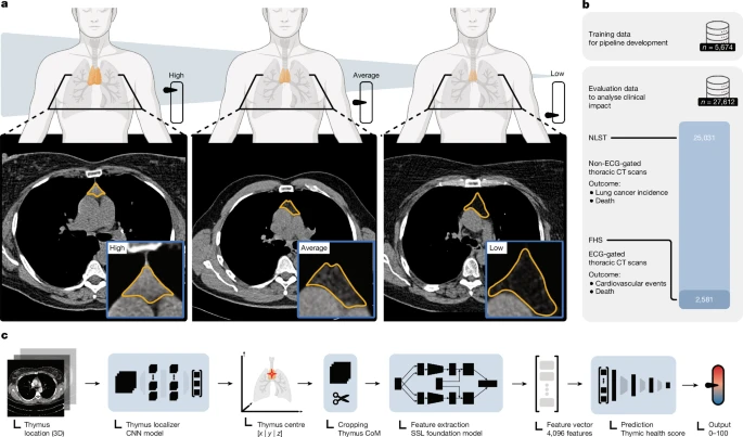
Fig 1 a, Illustration of thymic health, that is, an imaging-based proxy of thymic functionality and three representative examples of individuals with high, average or low thymic health. The thymus bed is outlined in orange. b, The model was developed on 5,674 CT scans and validated on 27,612 independent CT scans from the FHS and NLST. c, Illustration of the deep learning pipeline, which takes a CT scan as input and outputs a continuous quantification of thymic health after automatically localizing and quantifying the thymus on the basis of self-supervised learning (SSL). CNN, convolutional neural network; CoM, centre of mass; ECG, electrocardiogram. Illustrations in a and c created in BioRender; Birkbak, N. https://biorender.com/bd3dmmr (2026).
Fig. 2: Association of thymic health with long-term mortality.
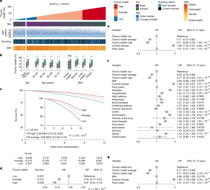
Fig 2 a, Overview of the NLST. Data are sorted by ascending thymic health where each column represents one patient. Thymic health is categorized into low, average and high based on the bottom 25% (blue), middle 50% (orange) and top 25% (red) of the population. The fractional left split of the NLST represents patients who were defined as having low thymic health through automatic quality control. b, Associations between thymic health and sex (n = 25,031) in the NLST, across age groups in years (n = 25,031) and body mass categories (n = 24,948, missingness n = 83). ***P < 2 × 10⁻¹⁶; NS, not significant with P = 0.2505. c, Kaplan–Meier plots for overall survival outcomes in the NLST across thymic health categories. The inset in the plot shows the same data on an expanded y axis. Unadjusted HRs are shown on the bottom left. d, HR for thymic health categories adjusted for pack-years and smoking status and stratified by sex and age binned at 5 years. e–g, HRs of all-cause death for participants in the NLST, using continuous age as the time scale to account for potential residual confounding by age (n = 25,027; missingness n = 4) adjusted for pack-years and smoking status and stratified by sex (e); adjusted for the shown clinical and epidemiological covariates and stratified by sex and age binned at 5 years (n = 24,597; missingness n = 434) (f); adjusted for the shown clinical and epidemiological covariates and stratified by sex and age binned at 5 years in the subgroup of the NLST in which participants with a history of cancer (n = 575), childhood or adult asthma, diabetes, asbestosis, bronchiectasis, lung fibrosis, sarcoidosis, silicosis or tuberculosis (n = 5,500) were excluded from the analysis (n = 18,565; missingness n = 54) (g). COPD, chronic obstructive pulmonary disease.
Thymic health and risk of death
To investigate the associations of thymic health with clinical outcomes, we assessed all-cause mortality throughout a 12-year follow-up period using Kaplan–Meier and Cox proportional hazards analysis. For the NLST, participants with higher thymic health showed lower mortality than those with low thymic health (high versus low thymic health: Kaplan–Meier mortality estimate at 12 years 13.4% versus 25.5%; hazard ratio (HR) 0.49; 95% confidence interval (CI) 0.45–0.53; Fig. 2c). The association was preserved in a Cox analysis adjusted for smoking status and pack-years, and stratified by sex and 5-year age bins (Fig. 2d). To verify that these results were not confounded by age, we repeated the analysis using narrower 3-year age bins (Supplementary Fig. 3a) and, in a separate model, used age as the time scale to implicitly adjust for the effect of ageing (Fig. 2e). To further evaluate the potential impact of comorbidities, we extensively adjusted for clinical variables and known diseases in a Cox model (Fig. 2f), and further repeated the analysis in a healthier sub-cohort where individuals with prior occurrence of cancer or major comorbidities were excluded (Fig. 2g). Throughout all analyses, thymic health remained significantly associated with outcomes after controlling for the potential confounding effects of either age or comorbidities (all type III P < 0.001), firmly indicating a prognostic value of thymic health beyond clinical variables. Finally, excluding thymic health from multivariate models did not markedly alter the HR estimates of the remaining covariates, indicating that thymic health provides independent and potentially complementary prognostic information (Extended Data Fig. 3a,e). These findings were supported in the FHS with similar effect size and direction (high versus low thymic health: 3.9% versus 14.5% Kaplan–Meier mortality estimate at 12 years; HR 0.24; 95% CI 0.16–0.38; average versus low thymic health: 9.6% versus 14.5%; HR 0.63; 95% CI 0.48–0.83; Extended Data Fig. 2c) (type III P < 0.001), although statistical significance was not reached after multivariate modelling in this smaller cohort (Extended Data Fig. 2d) (type III P = 0.254). Continuous and detailed cutoff analyses testing a wide range of thresholds confirmed these results, showing gradually improving survival independent of sex and age with increasing thymic health (Extended Data Fig. 1).
Lung cancer incidence and mortality
We investigated the associations of thymic health with lung cancer mortality and lung cancer incidence in the NLST, the main endpoints of the trial. Participants with high and average thymic health were less likely to develop lung cancer than participants with low thymic health (high versus low thymic health: 3.4% versus 5.3% Kaplan–Meier incidence estimate at 6 years; HR 0.64; 95% CI 0.53–0.76; average versus low thymic health: 4.1% versus 5.3%; HR 0.78; 95% CI 0.67–0.90; Fig. 3a). These associations were preserved in sex and age-stratified analyses with adjustments for smoking status and pack-years (type III P = 0.036) (Fig. 3b). Again, these associations were consistent across further analyses, extensively controlled for the confounding effects of age and comorbidities (Extended Data Figs. 3b,f, 4a,d and 5a and Supplementary Fig. 3b) (all type III P < 0.05). Furthermore, participants with higher thymic health had lower lung cancer mortality risks relative to participants with low thymic health (high versus low thymic health: 1.1% versus 2.0% Kaplan–Meier mortality estimate at 6 years; HR 0.52; 95% CI 0.44–0.63; average versus low thymic health: 1.5% versus 2.0%; HR 0.70; 95% CI 0.61–0.80; HR reported through 12 years; Fig. 3c). Again, these associations were preserved across the respective multivariate models (all type III P < 0.001) (Fig. 3d, Extended Data Figs. 3c,g, 4b,e and 5b and Supplementary Fig. 3c). We further stratified the cohort into current and former smokers. For lung cancer incidence, the associations with thymic health showed similar trends in both groups but did not reach statistical significance (current smokers type III P = 0.051; former smokers type III P = 0.25). For lung cancer mortality, the associations were preserved in both current (type III P = 0.009) and former smokers (type III P = 0.003) (Supplementary Fig. 4). While the NLST was designed to screen for lung cancer, pan-cancer mortality was also recorded (Supplementary Table 1). Again, we observed that participants with higher thymic health had lower cancer-specific mortality risks relative to participants with low thymic health, which were also preserved in multivariate models (all type III P < 0.02) (Extended Data Figs. 5c and 6a,b and Supplementary Fig. 3d).
Fig. 3: Association of thymic health with long-term risk of lung cancer and lung
cancer-specific mortality.
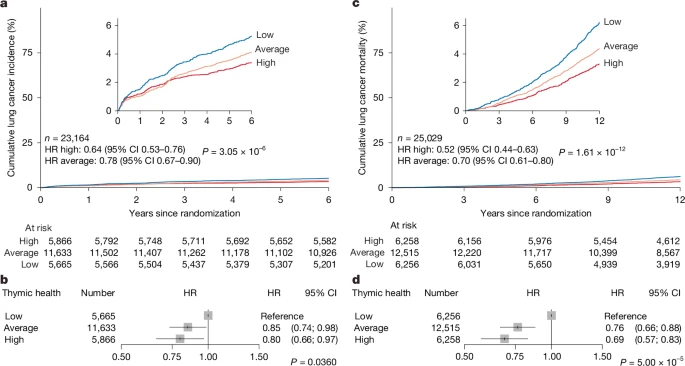
Fig 3a, Percentage of individuals who did develop lung cancer. b, HR of new lung cancer stratified by sex and age and adjusted for pack-years and smoking status. c, Lung cancer-specific mortality. d, HR of death from lung cancer stratified by sex and age and adjusted for pack-years and smoking status. The insets in the inverted Kaplan–Meier plots show the same data on an expanded y axis. a–d, Cox proportional hazards regression was used to estimate HRs. In the forest plots, the centre of each box represents the estimated HR, and the whiskers denote the corresponding 95% CI; shaded box size is for visualization only and does not encode statistical weight. The overall contribution of thymic health to uni- or multivariable models was evaluated using likelihood ratio tests (χ² tests) comparing full models with nested models excluding thymic health (type III test, two-sided) without adjustments for multiple comparisons.
Cardiovascular mortality and incidence
We investigated the associations of thymic health with cardiovascular-specific mortality in the NLST, where participants with high and average thymic health had lower cardiovascular-specific mortality risks than participants with low thymic health (high versus low thymic health: 2.9% versus 7.5% cardiovascular-specific mortality estimates at 12 years; HR 0.37; 95% CI 0.31–0.44; average versus low thymic health: 4.4% versus 7.5%; HR 0.57; 95% CI 0.50–0.65) (Fig. 4a and Supplementary Table 2). Again, these associations were preserved in sex and age-stratified analyses with adjustments for smoking status and pack-years (Fig. 4b) and also consistent across further sensitivity analyses, extensively controlled for the confounding effects of age and comorbidities (Extended Data Figs. 3d,h, 4c,f and 5d and Supplementary Fig. 3e) (all type III P < 0.001). Similar results were obtained in the independent FHS. Participants with higher thymic health had lower cardiovascular-specific mortality relative to those with low thymic health (high versus low thymic health: 0.3% versus 3.9% cardiovascular-specific mortality estimate at 12 years; HR 0.08; 95% CI 0.02–0.34, average versus low thymic health: 1.5% versus 3.9%; HR 0.38; 95% CI 0.20–0.70; Fig. 4c). These associations also were preserved in multivariate models (type III P = 0.021) (Fig. 4d). Further, participants with higher thymic health had lower cumulative incidence of cardiovascular-specific diseases relative to participants with low thymic health (high versus low thymic health: 5% versus 16.7% Kaplan–Meier estimate for cardiovascular disease at 12 years; HR 0.26; 95% CI 0.16–0.40, average versus low thymic health: 10.8% versus 16.7%; HR 0.65; 95% CI 0.49–0.87; Fig. 4e). However, these latter associations were partly attenuated in sex and age-stratified analyses with adjustment for smoking status (type III P = 0.051) (Fig. 4f).
Fig. 4: Association of thymic health with long-term risk of CVD-specific mortality,
CVD incidence and disease-specific mortalities.
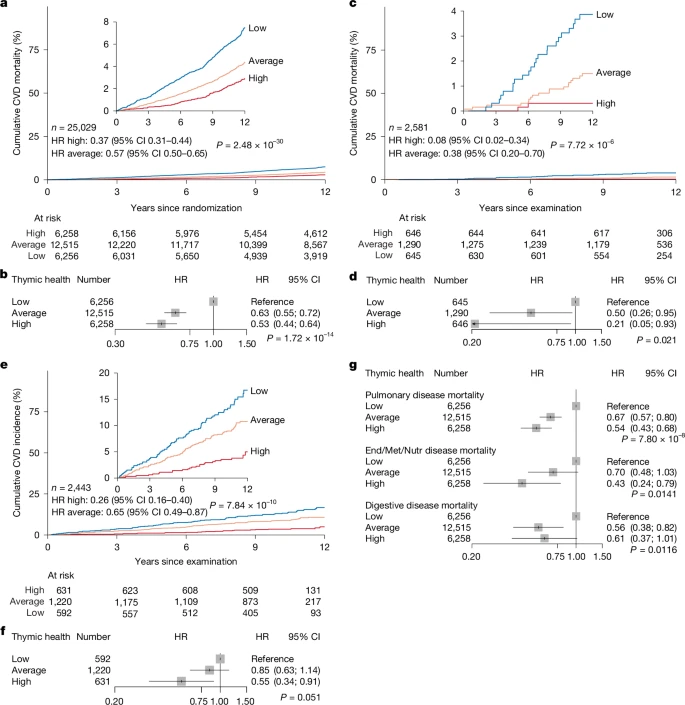
a, Percentage of participants in the NLST who died from CVD. b, HR of death from CVD adjusted for pack-years and smoking status, and stratified by sex and age. c, Percentage of participants in the FHS who died from CVD. d, HR of death from CVD adjusted for smoking status and stratified by sex and age. e, Percentage of participants in the FHS who had a new CVD-specific event, such as myocardial infarction, congestive heart failure or cerebral embolism. f, HR of a new CVD-specific event adjusted for smoking status and stratified by sex and age. g, HR of death from the specified disease groups among participants in the NLST stratified by sex and age and adjusted for pack-years and smoking status. a–g, Follow-up for all analyses was 12 years. The insets in the inverted Kaplan–Meier plots show the same data on an expanded y axis. Cox proportional hazards regression was used to estimate HRs. In the forest plots, the centre of each box represents the estimated HR, and the whiskers denote the corresponding 95% CI; arrowheads indicate that the 95% CI extends beyond the visualized limits; shaded box size is for visualization only and does not encode statistical weight. The overall contribution of thymic health to uni- or multivariable models was evaluated using likelihood ratio tests (χ² tests) comparing full models with nested models excluding thymic health (type III test, two-sided) without adjustments for multiple comparisons. End, endocrine; Met, metabolic; Nutr, nutritional.
Mortality by disease type
To investigate the relevance of thymic health in different disease types, aside from cancer and cardiovascular disease, we analysed the NLST, where the cause of death was recorded through a 12-year follow-up. Across all investigated disease-specific causes of death, participants with high or average thymic health were less likely to die as compared with participants with low thymic health (Extended Data Fig. 6), and all associations remained statistically significant in sex and age-stratified analyses with adjustments for smoking status and pack-years (Fig. 4g and Supplementary Fig. 3f) and in multivariate analyses using continuous age as the time scale to implicitly adjust for the effect of ageing (Extended Data Fig. 5e–g) (type III P < 0.05 for all). Mortality from pulmonary disease for high and average thymic health was 61% and 40% lower, respectively, compared to those with low thymic health (Extended Data Fig. 6c and Supplementary Table 3). Likewise, mortality from endocrine, nutritional and metabolic diseases, including metabolic disorders such as diabetes mellitus, was 68% and 37% lower for individuals with high and average thymic health, respectively, compared to those with low thymic health (Extended Data Fig. 6d and Supplementary Table 4). Finally, mortality from diseases of the digestive system that included liver, gallbladder or pancreatic diseases was 54% and 47% lower for individuals with high and average thymic health, respectively, compared to those with low thymic health (Extended Data Fig. 6e and Supplementary Table 5). Together, these data provide evidence that thymic health is prognostic across diverse diseases, indicating disease-agnostic relevance for health.
Impact of metabolic health and lifestyle
Next, we investigated the associations of cholesterol, triglycerides, fasting glucose and blood pressure with thymic health in the FHS (Fig. 5). Across all clinically relevant categories, female participants had higher thymic health as compared with male participants, and thymic health was positively associated with metabolic health (Fig. 5a,b). In sex-, age- and smoking status-adjusted analyses, and across sex strata, we consistently found that high-density lipoprotein (HDL) had a significant positive association with thymic health, while the common variables of metabolic syndrome, including triglyceride levels, fasting glucose and blood pressure, showed negative associations with thymic health (Fig. 5b and Supplementary Fig. 5).
Fig. 5: Associations of thymic health with metabolism, lifestyle and inflammation in
the FHS.
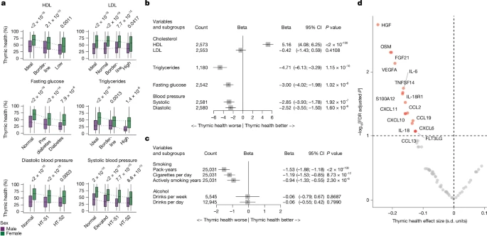
a, Associations of metabolically relevant variables with thymic health, stratified by sex with female participants shown in green and male participants in purple. b, Respective associations adjusted for sex, age and smoking status. c, Associations of smoking-related factors and weekly consumption of alcoholic beverages with thymic health adjusted for sex and age. d, Association of Olink-based plasma protein levels with thymic health, adjusted for sex, age and smoking status (n = 317). In a, box plots show the median (centre line), interquartile range (25th–75th percentiles; box), and whiskers extending to the minimum and maximum values within 1.5× the interquartile range. Statistical comparisons between male and female participants were performed using two-sided Wilcoxon rank sum tests. Patient counts shown in a correspond to the number (Count) of patients depicted in the respective rows of b. In b and c forest plots, the box centres represent the estimated regression coefficients, and the whiskers their corresponding 95% CI. The shaded box size is for visualization only and does not encode statistical weight. Statistical significance of individual coefficients was evaluated using two-sided t-tests. In a–c no adjustment for multiple comparisons was applied. In d, effect size, in s.d. units, is plotted against false discovery rate (FDR) of the linear regression association of rank-based inverse-normal transformed Olink inflammatory proteins with thymic health. The FDRs were computed across 68 proteins using the two-sided regression P values using the Benjamini and Hochberg method for multiple comparisons. Proteins with FDR < 0.1 (corresponding to −log10[FDR] > 1) were considered statistically significant. HT-S1, hypertension stage 1; HT-S2, hypertension stage 2; LDL, low-density lipoprotein.
Furthermore, we used the Fried frailty phenotype23 to investigate whether individuals with low thymic health are at increased risk for future disabilities and reduced quality of life. We found a significant association between low thymic health and increased frailty scores (P < 0.001), independent of sex, age and smoking status (Supplementary Table 6). Exploring the individual components of the Fried frailty index, we found that this signal was particularly driven by slower walking speed (P = 0.032), lower physical activity index (P = 0.010) and increased exhaustion (P = 0.021). Together, these data indicate a strong relationship between metabolic health and lifestyle with thymic health, and that thymic health may directly be associated with an individual’s quality of life and long-term risk of disability.
Chronic inflammation and thymic health
As immune response and inflammation are closely related, we explored whether low thymic health is associated with dysregulated inflammatory processes. For this, we investigated the association of blood inflammatory proteins with thymic health. First, we analysed proteomic blood plasma data obtained from 317 individuals included in FHS (Supplementary Table 7) before the CT scans (mean time difference, 10.4 years). Plasma samples were analysed using the Olink inflammation panel comprising 92 proteins, with 68 passing quality control. Of these, 16 out of 68 (24%) proteins were negatively associated with thymic health (Fig. 5d), including major mediators and regulators of inflammation, such as vascular endothelial growth factor A (VEGFA), interleukin-6 (IL-6), IL-18, hepatocyte growth factor (HGF), oncostatin M (OSM) and C-X-C motif chemokine ligand (CXCL) family members 10 and 11. These results show that individuals with higher systemic levels of inflammation had lower thymic health, indicating an interplay between inflammatory processes and thymic health.
To investigate this further, we assessed whether chronic inflammation lasting for a prolonged period of time correlated with thymic health. For this, we collected longitudinal C-reactive protein (CRP) measures over 5 to 10 years for 1,156 participants in the FHS (Supplementary Table 8). We found that participants in the FHS with chronic systemic inflammation, quantified by consistently high CRP levels of greater than or equal to 3 mg l−1 (140 out of 1,156, 12.1%) over multiple longitudinal blood measurements, had substantially lower thymic health independent of sex, age and smoking status (P = 0.0012).
Taken together, these data highlight that inflammation and thymic health are closely related; one potentially driving and potentiating the other, with associated negative health consequences.
Thymic health model stability
To assess model stability, we performed stability investigations that demonstrated excellent test–retest stability and robustness to inter-reader input variations (Extended Data Fig. 7). Furthermore, to investigate which anatomical regions the model relies on for making its quantifications, we performed activation mapping, which demonstrated specific attention of the model to the thymic bed, while giving minor attention to adjacent structures, indicating knowledge of the anatomical context (Extended Data Fig. 8). Taken together, these results demonstrate that the performance of the thymic health model is robust against input variations, and that it captures contextual information directly from the anatomical region of the thymus. Extensive details regarding model development and testing can be found in Supplementary Methods S1 and S2, Supplementary Figs. 9–17 and Supplementary Tables 9–13.
Discussion
Our results show that thymic decay in adults is highly individualized and that loss of thymic health increases mortality and disease incidence, including cancer and cardiovascular diseases. These findings were investigated in two independent prospectively collected clinical studies of asymptomatic adults, the NLST and the FHS. Among more than 25,000 participants in the NLST, higher thymic health was consistently associated with a significantly lower risk of mortality by any cause, lung cancer and cardiovascular diseases, independent of sex, age, smoking, prior diseases or cancer history. Similar results were found in the independent FHS, where participants with high thymic health had a significantly reduced risk of death from cardiovascular disease, a primary endpoint of the study, independent of sex, age and smoking. Considering the prevailing perception of the limited role of the thymus in adults, our findings may change our understanding of human health, emphasizing the complex yet critical role of the immune system in long-term well-being and longevity.
We found that thymic health was associated with critical health consequences. Presumed healthy participants in the NLST with high thymic health had an approximately 50% reduction in the risk of death, were 36% less likely to develop lung cancer and nearly 50% less likely to die from lung cancer, as compared to participants with low thymic health. Further, the significant impact of high thymic health on cardiovascular disease (CVD) was consistent across participants in the NLST and FHS, with risk reductions in CVD mortality ranging from 63% to 92%. Given the critical role of the thymus in generating a diverse T cell repertoire24, these results support the broad impact of a sustained adaptive immune system to combat disease and promote longevity.
Our results challenge the established notion that the cessation of thymic output in ageing adults is inconsequential as it is naturally replaced by peripheral expansion of T cells7. Rather, our results suggest that loss of thymic tissue in adults may forecast higher risks of disease and death. The main function of the thymus is to generate a diverse T cell repertoire, which provides adaptive immunity throughout life7. While the relevance and abundance of the T cell repertoire at a young age are well documented7, our results indicate that the thymus retains a continued role in T cell production throughout adulthood and that the pattern of decline of thymic function in adults is associated with poorer health outcomes.
A recent landmark study demonstrated an association between thymectomy and reduced lifespan and increased risk of cancer, among other effects8. While thymectomy is rare, we demonstrate that the thymic decay is highly individualized even in presumed healthy adults, indicating that thymic function can also be substantially reduced in individuals who did not have their thymus surgically removed. Our work therefore impacts the wider population and aligns with the well-established age-related decline in immune system function6,25,26. This is also consistent with previous work modelling declining T cell output as a major risk factor for age-related increase of cancer development13.
Lifestyle and metabolic health measures, such as smoking, physical activity or HDL levels, showed strong associations with thymic health. Likewise, in longitudinal blood-based evaluations, we found individuals with chronic inflammation, a hallmark of immunosenescence and commonly associated with chronic stress, carbohydrate-rich diet and obesity, had lower thymic health.
Among presumed healthy individuals from the FHS, lower thymic health was indeed associated with pro-inflammatory modifications of blood plasma protein levels, consistent with the presence of chronic inflammation. The pro-inflammatory pattern included increased levels of cytokines IL-6, IL-18 and OSM, as well as several CXCL chemokines, all of known relevance in systemic inflammatory diseases such as atherosclerosis, age-associated diseases such as arthritis, and cancer27.
Our findings are further supported by studies in which thymic involution was associated with immunosenescence and inflammation, contributing to illnesses such as metabolic or cardiovascular disease12. Metabolic syndrome affects more than a third of all adults in the USA, with continuously increasing prevalence28. Our results demonstrate significant associations between metabolic and thymic health. These findings are consistent with those of recent studies in which fatty degeneration of the thymus was associated with obesity18,19,20 and smoked pack-years19,20. Together, these findings suggest a profound impact of actionable lifestyle choices on thymic health and may further clarify why healthy behaviour improves well-being and lifespan.
In addition, the global increase in early-onset cancers might be linked to an accelerated rate of thymic decay, potentially driven by factors such as smoking, minimal physical activity and overall unhealthy lifestyles, leading to more inflammation in the body. Indeed, thymic health could refine disease and cancer screening strategies, especially for high-risk individuals.
Here we examined the impact of thymic health on lung cancer incidence separately in current and former smokers. Associations were primarily driven by current smokers, likely reflecting both higher statistical power due to higher event rates and the greater biological relevance of thymic function under ongoing tobacco exposure and thereby continuously increasing neoantigen load, making thymic health particularly relevant for immune surveillance in this setting29.
Previous studies have attempted to quantify thymic characteristics using imaging techniques, using conventional approaches such as estimating the proportion of fatty degeneration or measuring attenuation density18,19,20. However, these studies were limited in scope, focusing on the exploration of associations between thymic features and basic clinical and epidemiological measures. Previous studies did not find any associations between thymic imaging characteristics and outcomes such as survival or disease incidence. These previous investigations reported presumed residual thymic tissue in only a minor fraction of the examined population ranging from 41%18 to 26%19,20, that is, the studies estimated a fully fatty degenerated thymus in approximately 60–75% of individuals18. We find these results contradictory to biological associations of sustained T cell output in visually fully fatty degenerated thymic glands18, and we could corroborate that our measure of thymic health had preserved impact on outcomes across individuals who had no appreciable visual thymic tissue according to independent visual scoring, indicating insufficient thymic quantification using a visual scoring system. Our deep learning model found favourable thymic functionality among individuals with average and high thymic health, representing 75% of participants, consistent across both independent study populations and paralleled by substantial health benefits in these participants. Indeed, our findings are supported by Kooshesh et al.8, who found substantial negative health consequences after thymectomy in adults, indicating relevant thymic activity throughout life across the population.
Participants included in this study were prospectively enrolled in the FHS and NLST, covering a wide age range for both sexes. They are, however, predominantly white, and further validation in more ethnically diverse populations is warranted. While we observed an association between thymic health and overall survival in both cohorts, this did not remain significant following thorough multivariate adjustment in the smaller FHS. This further indicates that long-term outcomes may be influenced by dynamic lifestyle choices in this population that benefits from regular medical examinations and follow-up, and supports the notion that active engagement in health-promoting behaviours may potentially attenuate the long-term negative impact of unhealthy characteristics associated with low thymic health30. Taken together, our results provide evidence of multifaceted associations between thymic health and clinically relevant health consequences across clinical, epidemiological and biological characteristics and strongly suggest the critical relevance of the thymus for adult health.
Before the thymic health deep learning model developed here can be applied in a clinical setting, it is essential to prove generalizability. Development and application of the thymic health model were performed in fully independent datasets, with high robustness as demonstrated by test–retest stability. Given substantial differences in acquisition protocols, scanners and population characteristics between the FHS and the NLST, the thymic health analyses were conducted using population-specific thresholding, and no universal cutoffs can be assumed. Future studies with international and external validation are needed to explicitly address batch and scanner variability and enable direct cross-cohort comparisons and generalizable thresholds.
Our results connect thymic health with longevity and lower disease incidence. Our results suggest that inflammation potentially drives an accelerated rate of thymic decay. This finding may provide new opportunities for preventive strategies that aim to reduce thymic decay and potentially even reverse it. These can include the use of anti-inflammatory and anti-obesity drugs and open new avenues of drug development that aim to improve long-term immune health. Furthermore, even practical approaches, such as lifestyle changes including exercise and sleep, as well as healthy food choices and supplement intake, are likely to notably impact thymic health.
While this study investigated the role of thymic health in healthy individuals, the state of the immune system could also have an important role in individuals with disease. This is particularly relevant for treatments that rely on triggering an immune response, such as immunotherapies used to treat patients with cancer. However, it may also be relevant for other diseases: for example, during the recent COVID-19 pandemic, the response to the virus varied significantly between sex and age groups, with older men particularly affected31.
Our analysis reveals previously unrecognized, possible negative consequences of reduced thymic health. While thymic health declines with age, we also find considerable variation in thymic health within age groups, indicating that the rate of decay varies considerably between individuals. When we analysed blood inflammatory proteins, we found an association between increased levels of inflammatory proteins and thymic decay. Although this analysis was limited by a relatively long interval of 10.4 years between the CT scans used for thymic health assessment and blood samples used to assess protein content, these results were supported by an orthogonal assessment of CRP levels, supporting that chronic inflammation may be a likely driver of poor thymic health. It is also likely that a genetic component exists, predisposing certain individuals to increased or decreased rates of thymic decay. A recent large-scale study highlighted substantial individual variability in immune resilience and its link to long-term health outcomes32. Gene expression profiles associated with immune competence and low inflammation were linked to longevity, while pro-inflammatory signatures correlated with poorer outcomes. These findings align with our observations that thymic health varies widely across individuals and is negatively associated with chronic inflammation. Awareness of a genetic predisposition may lead to preventive measures and increased surveillance, and this should be elucidated in future studies that match single nucleotide polymorphisms with rates of thymic decay. Other limitations of this study include the older age and heavy smoking status of individuals in the NLST cohort relative to the FHS cohort, both likely to affect thymic health.
An important implication of our study is that the retrospective observational design does not allow conclusions about causality. It is possible that lower thymic health contributes to adverse outcomes by weakening immune resilience, but it is also possible that pathological processes leading to reduced health status, mortality and disease drive thymic decline. Clarifying the direction and nature of these associations will require future prospective and mechanistic studies and will be essential to determine whether thymic health can serve as a target for prevention or intervention strategies. Recent large-scale initiatives, such as the Advanced Research Projects Agency for Health-funded Thymus Rejuvenation programme, underscore growing interest in developing regenerative approaches to restore thymic function, further highlighting the high clinical relevance of this work33.
In summary, this study underscores the highly personalized nature of thymic health and emphasizes the previously unrecognized possible critical role of maintaining thymic health to preserve an agile, adaptive immune response that will accommodate long-term well-being and longevity. Today, thymic assessments do not have an established clinical standard, and the thymus is not examined in routine clinical care. The extent to which the adult appearance of the thymus is associated with health and whether actionable lifestyle or risk factors may be harnessed to improve thymic health was unknown. By analysing 27,612 individuals, our results provide evidence that thymic health is directly associated with critical outcomes and diseases and may be directly targetable by various approaches, such as smoking reduction and weight loss in overweight and obese individuals.
Our results underline the relevance of the thymus throughout life. Clinical investigations of preventive or regenerative strategies will be vital to help us understand how to harness the thymic potential to improve population health34,35,36,37,38.
READ ORIGINAL ARTICLE FOR METHODS AND REFERENCES