The Role of Gut Microbiome-Derived Short-Chain Fatty Acid Butyrate in Hepatobiliary Diseases

Kishor Pant∗ Send email to kpant@umn.edu ∙ Senthil K. Venugopal† ∙ Maria J. Lorenzo Pisarello‡,§ ∙ Sergio A. Gradilone∗, Send email to sgradilo@umn.edu

Abstract

The short-chain fatty acid butyrate, produced from fermentable carbohydrates by gut microbiota in the colon, has multiple beneficial effects on human health. At the intestinal level, butyrate regulates metabolism, helps in the transepithelial transport of fluids, inhibits inflammation, and induces the epithelial defense barrier. The liver receives a large amount of short-chain fatty acids via the blood flowing from the gut via the portal vein. Butyrate helps prevent nonalcoholic fatty liver disease, nonalcoholic steatohepatitis, inflammation, cancer, and liver injuries. It ameliorates metabolic diseases, including insulin resistance and obesity, and plays a direct role in preventing fatty liver diseases. Butyrate has different mechanisms of action, including strong regulatory effects on the expression of many genes by inhibiting the histone deacetylases and modulating cellular metabolism. The present review highlights the wide range of beneficial therapeutic and unfavorable adverse effects of butyrate, with a high potential for clinically important uses in several liver diseases.
Liver, the largest internal organ of the human body, plays a role in many physiological functions including glycogen, protein, and enzyme synthesis, and can self-regenerate. It also metabolizes the body’s harmful toxins, which involves well-controlled biochemical processes., Increasing evidence suggests that butyrate, a gut microbiota-derived short-chain fatty acid (SCFA), could be helpful in the prevention of liver diseases and metabolism regulation. SCFAs in the liver help in many aspects of liver disease, including steatosis, hepatitis, inflammation, and hepatocellular carcinoma (HCC). Butyrate is the most important SCFA for many bodily functions, including those in the colon and liver. This review highlights the critical functions of SCFAs, particularly those of butyrate in normal physiology and its prospective therapeutic effects in many liver diseases.
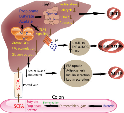
Butyrate is composed of four carbon atoms and originates in the intestine from the bacterial fermentation of plant-origin food materials, such as fibers, celluloses, and complex sugars; mammals lack the necessary enzymes to metabolize these compounds. Acetic, propionic, and butyric acids are the predominant forms of SCFAs in the animal body. SCFAs have several positive effects in animals (Figure 1). For ruminants and herbivorous animals, SCFAs are the primary substrates for energy production.

Figure 1. Short-chain fatty acids (SCFAs) produced in the colon by gut microbiota travel mainly to the liver via the portal vein and the various parts of the body. They primarily help in the inhibition of nonalcoholic fatty liver disease (NAFLD) by lessening the process of lipogenesis, free fatty acid (FFA) accumulation, and glucose output. Furthermore, SCFAs also inhibit lipopolysaccharide-induced inflammation as they alleviate IL-1, tumor necrosis factor-α (TNF-α), cyclooxygenase-2 (COX-2), and inducible nitric oxide synthase (iNOS) production in the liver. Butyrate inhibits cell proliferation and cell cycle progression and induces apoptosis in cancer cells. HCC, hepatocellular carcinoma; HDAC, histone deacetylase; TG, triglycerides; TLR4, toll-like receptor 4.

Butyric acid has received more focus among all three SCFAs, and it is a natural substance in most animals’ gut, milk, and feces. Butyrate is naturally present in high concentrations in the gut and large intestine. Although previous investigations suggested its role as a growth factor for normal cells, recent studies have focused more on the antitumor properties of butyrate.,

Gut-Liver Axis and the Metabolism of Butyrate
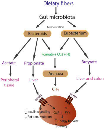
The gut microbiota, mainly present in the large intestine region, are essential in providing energy to the host by producing SCFAs via anaerobic fermentation (Figure 2). Complex polysaccharides, oligosaccharides, and oligofructose are resistant to the hydrolytic enzymes in the human gut. Hydrolytic enzymes from the gut microbiota, including β-fructosidase, β-galactosidase, and xylanase, are mainly used to ferment these molecules. During fermentation, energy is acquired as ATP by substrate-level phosphorylation through substrate breakdown via oxidation. This process also occurs in oxygen-deprived conditions and leads to reduced end products that can act as terminal electron acceptors, such as butyrate.

Figure 2. The illustration shows the formation of the short-chain fatty acids, including acetate, propionate, and butyrate, in the colon by the gut microbiota during the fermentation of the dietary fibers. Bacteroids produce propionate and acetate, whereas Eubacteria is responsible for butyrate formation. Butyrate activates G-protein receptor-41 (GPR41) to activate glucagon-like peptide-1 (GLP-1) and peptide YY (PYY), thereby increasing the energy harvest and satiety, which eventually may decrease fat accumulation in the body.
The fermentation of these carbohydrates in the gut is mainly assisted by the Bifidobacteria, Lactobacilli, Flavobacterium, Bacteroides, and a group of Gram-negative bacteria, Clostridium. It is well established that butyrate-producing bacteria contribute an essential function in sustaining gut health, mainly via the generation of butyrate. This short-chain fatty acid stabilizes the intestinal lumen pH, which is necessary for proper digestion and absorption of nutrients. Moreover, several species of butyrate-producing bacteria have shown anti-inflammatory activities, which may help lower gut inflammation and boost overall gut health. One of the most critical advantages of butyrate-producing bacteria is their capacity to be tolerated by the innate immune system. This indicates that the body’s natural defense system recognizes and accepts these bacteria, which is crucial for maintaining a healthy gut microbiome.
The gut and liver are connected through the portal vein. Through this connection, the liver can be affected by the intestinal microbiota and its metabolites, forming the gut-liver axis. Butyrate is mainly taken up by the epithelium cells of the colon and metabolized to produce energy; however, the blood flowing through the colon area also carries butyrate to many parts of the body, including the liver.
Once produced, the SCFAs may be absorbed into the circulation via the intestinal lining. Specialized transporters, like monocarboxylate transporter 1 (MCT1) (SLC16A1) and monocarboxylate transporter 4 (MCT4) (SLC16A3), are H+-coupled transporters that facilitate the electroneutral transport of SCFAs and are located on the epithelial cells that line the colon and enable the absorption of SCFAs. These transporters move SCFAs across the cell membrane and into circulation, where they can be delivered to various tissues and organs. SCFAs, including butyrate, are
transported to the liver via the hepatic portal vein.,
Once in the liver, SCFAs may be metabolized by liver cells and converted to several other molecules, including glucose, ketone bodies, and fatty acids, which other tissues and organs can then use. The surplus SCFAs enter the circulation and are transferred to several tissues and organs throughout the body, including adipose tissue, muscle, and the brain. Nevertheless, the transportation of SCFAs from the liver to other organs is not well known and may include numerous pathways, such as transporters and membrane diffusion.
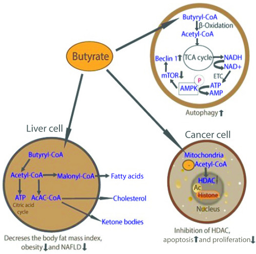
In the mitochondria of liver cells, butyrate undergoes β-oxidation, a process that breaks down fatty acids to produce acetyl-CoA. The enzyme butyryl-CoA dehydrogenase converts butyrate to butyryl-CoA, which is subsequently converted to acetyl-CoA by the enzyme butyryl-CoA synthase. The acetyl-CoA then enters the citric acid cycle to produce ATP energy (Figure 3). Butyrate metabolism in the liver also helps other metabolic processes and energy generation. Some of the acetyl-CoA generated from butyrate, for instance, may be used to synthesize cholesterol and other lipids. Butyrate also accelerates gluconeogenesis, the production of glucose from noncarbohydrate sources, such as amino acids and fatty acids. This is achieved by converting acetyl-CoA to pyruvate, which may subsequently be used in gluconeogenesis to produce glucose. The liver absorbs the glucose for energy production or stores it as glycogen for later use, providing approximately 30% of the hepatic energy requirements.

Figure 3. Diverse effects of butyrate in the body are shown. Butyrate is an energy source converted to acetyl-CoA, which enters the tricarboxylic acid (TCA) cycle in mitochondria. It inhibits histone deacetylases (HDACs), inhibiting cell proliferation and cell cycle, and up-regulates apoptosis and autophagy in cancer cells. AMPK, AMP-activated protein kinase; ETC, electron transport chain; mTOR, mammalian target of rapamycin; NAFLD, nonalcoholic fatty liver disease.


Three primary SCFAs are produced by the gut microbiota during fiber fermentation: acetic acid, propionate, and butyrate. Although these three SCFAs are the most well known and studied, other minor SCFAs, such as formate, valerate, and isovalerate, can also be produced in smaller amounts. However, their contribution to physiological processes and health outcomes is not as well established as that of the main SCFAs. The composition and quantities of SCFAs produced can vary, depending on factors such as dietary fiber, gut microbiota, and individual differences.,
In the human colon, butyrate concentrations typically range from 10 to 20 mmol/kg of feces or approximately 5 to 20 mmol/L in the gastrointestinal tract. Acetate and propionate, the other two major SCFAs, are usually present in higher concentrations than butyrate. Acetate concentrations can range from 70 to 140 mmol/kg of feces or approximately 60 to 140 mmol/L in the gut. Propionate concentrations can range from 10 to 30 mmol/kg of feces or approximately 5 to 20 mmol/L in the gastrointestinal tract. These are approximate ranges and can vary among individuals., The relative concentrations of SCFAs depend on factors such as dietary fiber, gut microbiota, the metabolic activities of specific bacterial strains in the colon, and individual genetics.,
In the liver, butyrate concentrations are typically in the micromolar range. Studies have reported hepatic butyrate concentrations in human liver samples ranging from approximately 10 to 100 μmol/L. Acetate concentrations in the liver can range from approximately 100 to 500 μmol/L, whereas propionate concentrations are typically lower than acetate, ranging from approximately 10 to 100 μmol/L in human liver samples.,

Role and Beneficial Properties of Butyrate

Although butyrate, propionate, and acetate have important roles in cellular functions, the precise effects and mechanisms of action could vary. The following butyrate properties are the reason it is often considered to be more effective than propionate and acetate in some biological processes. First, butyrate is a more potent inhibitor of histone deacetylases (HDACs). The median inhibitory concentration required to inhibit HDAC activity was measured to be 223 ± 64 μmol/L for propionate, whereas for butyrate it was found to be 52 ± 11 μmol/L., Second, studies have demonstrated that butyrate facilitates cellular differentiation. Last, butyrate exhibits strong anti-inflammatory effects., Comparatively, although propionate and acetate also have cellular effects, in certain conditions they may not be as potent as those of butyrate, mostly because of butyrate’s anti-HDAC activity and anti-inflammatory activity at lower concentrations.,

Immunomodulatory Effects of Butyrate

An interaction between microbiota and host is essential for the active function of the immune system. The development of immune cells, production of antibodies, and production of antimicrobial peptides in the host are modulated by the microbiota and its metabolites. The loss of equilibrium between the immune system and microbiota is linked to the development of inflammatory bowel disease (IBD), rheumatoid arthritis, diabetes, and obesity in the host.
Butyrate augments the expression of antimicrobial peptides by epithelial cells and modulates the assembly of immune mediators and cytokines, like IL-18, required to restore and protect epithelial integrity. SCFAs also regulate the activation, recruitment, and differentiation of immune cells, including dendritic cells, neutrophils, macrophages, and T lymphocytes. SCFAs modulate neutrophils and activate the effector function and survival in the tissues. In addition, SCFAs induce direct effects in T-lymphocyte proliferation and differentiation and in the generation of T-regulatory cells. Type 1 helper T cells use butyrate as an energy factor for the cells. However, in the presence of antigen-presenting cells, butyrate inhibits type 1 helper T cell proliferation and blocks lymphocyte proliferation. In human lymphocytes, butyrate induces the secretion of IL-10 and IL-2.,
IBD is a recurrent chronic gastrointestinal tract disorder related to the immune system. Hepatobiliary disorder manifestations are widespread in IBD and have various etiopathogenetic factors. IBD can develop active inflammation in the liver if it remains untreated. In a study by Bailón et al, butyrate at low concentration inhibited the proliferation of immune cells and the activation state of cells involved in IBD. Butyrate also induced apoptosis in activated T cells by caspase-3/7 activation and secretion of inflammatory cytokines. The inflammatory cells generate significant reactive oxygen species in the cells that induce colorectal cancer. Butyrate ameliorates the hydrogen peroxide–induced injury in colon cells, modulating the gene expression related to oxidative stress., In human subjects, the rectal administration of butyrate enhanced the antioxidant capacity in the colon.
Butyrate treatment in high-fat diet fed mice reduces liver steatosis and inflammation. Treatment with butyrate significantly reduced tumor necrosis factor (TNF)-α expression in the liver and restored glucose transporter (GLUT) and peroxisome proliferator-activated receptor gene expression in the liver and adipose tissue. Butyrate may have the potency to regulate proinflammatory cytokines and genes in the liver via inhibition of toll-like receptors (TLRs) and NF-κB activation. Patients with IBD have a low level of butyrate-producing bacteria and decreased butyrate transporters in the colonic mucosa, which is responsible for IBD pathogenesis., Twenty healthy young individuals were randomly assigned to a high-SCFA diet or a low-SCFA diet for 21 days each, with a 21-day rest interval in between, to assess the effects of a high-SCFA diet on colonic fermentation and immune cells. High-SCFA diet led to higher fecal and plasma SCFA concentrations and reduced fecal ammonia concentrations. It also resulted in significantly lower blood total B cells, naive B cells, type 1 helper T cells, and mucosal-associated invariant T cells than the low-SCFA diet. Therefore, interventions to increase colonic and peripheral blood butyrate may affect circulating immune cells in humans. However, whether these changes have immunomodulatory effects and are therapeutically promising warrants further investigations.
In a study by Säemann et al, butyrate demonstrated anti-inflammatory properties via inhibition of IL-12 and up-regulation of IL-10, which may have some implications in the modulation of immune responses. A study where 4 g of sodium butyrate was given daily for 4 weeks to 9 healthy and 10 obese individuals was performed to assess the impact on cytokine responses and the induction of trained immunity. Butyrate supplementation had a considerable impact on trained immunity in the monocytes of obese people with metabolic disorders, reducing their overall inflammatory status while having only little impact on the direct stimulation of cytokine production. Finally, other studies show that oral butyrate supplementation may help patients with metabolic syndrome reduce inflammation.,
In a co-culture system (differentiated 3 T3-L1 adipocytes and RAW264.7 cells), butyrate significantly inhibited inflammatory cytokines (TNF-α, monocyte chemoattractant protein-1, and IL-6), free glycerol, and free fatty acids. Furthermore, butyrate inhibited mitogen-activated protein kinase phosphorylation, NF-κB activity in cultured macrophages, and lipase activity in adipocytes. In patients with IBD, butyrate treatment decreased the production of inflammatory cytokines (TNF-α, IL-1β, and IL-6),, and inhibited inflammation via inhibition of TNF-α and NF-κB expression, thereby reducing pathogenesis of Crohn diseases in the patients. Butyrate inhibits the production of lipopolysaccharide-induced inflammatory cytokines (TNF-α and interferon-γ), NF-κB, inducible nitric oxide synthase, and nitric oxide from cultured neutrophils and cells., In addition to controlling cytokine production, reduction in SCFA-induced luminal pH also prevents harmful microorganisms from proliferating. The antimicrobial protein cathelicidin IL-37 is induced by SCFAs, particularly butyrate, and increases the number of T-regulatory cells in the gut, which both support host defense.

Role of Butyrate in Liver Diseases

Alcoholic Liver Disease

Alcohol consumption leads to intestinal dysbiosis and changes in SCFA concentrations. Administration of probiotic strains, such as Pediococcus pentosaceus, has been shown to counteract the decrease of butyrate in animal studies. Treatment with P. pentosaceus reduces endotoxin levels and ameliorates inflammation and alcohol-induced hepatic steatosis.
Oral supplementation of a symbiotic agent, a combination of a butyrate-producing bacteria and a butyrate-yielding prebiotic, in mice with liver damage due to chronic alcohol consumption showed increased butyrate transporters in the liver and consequent improvement of inflammation and oxidative stress.
Nonalcoholic Fatty Liver Disease

Nonalcoholic fatty liver disease (NAFLD) is a common liver ailment and a significant public health problem worldwide. NAFLD starts with simple fat deposition (steatosis) but may further develop into nonalcoholic steatohepatitis (NASH), fibrosis, and HCC., Administration of a high-fat diet in mice decreases butyrate formation and increases liver fat content and inflammation. However, the presence of fiber in the mice fed with a high-fat diet reduces weight gain, liver fat content, triglyceride, and cholesterol levels in the body and increases the formation of butyrate. Butyrate-producing probiotic (MIYAIRI 588) prevents NAFLD progression by enhancing the expression of AMP-activated protein kinase (AMPK), AKT, and nuclear factor erythroid 2–related factor 2 (Nrf2) in mice.
Fibroblast growth factor 21 (FGF21) is abundantly expressed in the liver, and reduces fat deposition via inducing lipid oxidation, energy expenditure, and ketogenesis. FGF21 is known to be down-regulated in NAFLD and obesity in human and nonhuman primates., Studies exploring the role of FGF21 in the metabolism of butyrate indicate that FGF21 is activated by butyrate and stimulates fatty acid oxidation in the liver. Butyrate also stimulates FGF21 transcription by inhibiting HDAC3 expression.
Diabetes mellitus or insulin resistance deregulates the blood glucose and lipid level in patients and contributes to the development of NAFLD in humans., Butyrate can augment glycogen synthesis at a rate similar to glucose in the liver. Therefore, consuming fiber may prevent insulin resistance in humans via the production of butyrate. An experimental diet fortified with butyrate was shown to ameliorate diabetes by reducing insulin resistance. A study by Henagan et al demonstrated that butyrate treatment effectively improves body fat level and glucose tolerance in obese mice by involving nuclear-encoding mitochondrial gene expression regulation through a particular nucleosome repositioning, which resulted in improved mitochondrial β-oxidation. Overall, these studies show that dietary butyrate supplementation prevented diet-induced insulin resistance in mice by inducing energy expenditure and mitochondria function.
Obesity is another condition where body fat or mass increases may also contribute to the development of NAFLD., Supplementation of butyrate by butyrogenic food (dietary fiber) effectively lowered body fat and helped treat obesity in obese mice., Gut microbiota produces 50 to 100 mmol/L per day of SCFAs, including propionic and butyric acid as main carbohydrate fermentation products. SCFAs elicit their effects by binding to the G-protein–coupled receptors (41 or 43) on the gut endocrine cells, immune system, and adipocytes. A diet containing sugar-beet fibers produced SCFAs via cecal fermentation and reduced hepatic cholesterol synthesis and fat content compared with rats on a fiber-free control diet. In another study, SCFA (butyrate and propionate, 5% by weight) consumption lowered hepatic fatty acid synthase activity and hepatic fat and increased the level of hepatic lipid oxidation in high-fat (45%) fed mice.
A recent investigation looking at butyrate’s positive benefits found that it may be used therapeutically to treat NAFLD and other related metabolic disorders. In the study, a mouse model of NAFLD was induced by feeding mice a high-fat diet and giving them sodium butyrate for 8 weeks. In mice fed a high-fat diet, butyrate increased the activity of genes triggered by insulin and inhibited genes that are lipogenic, which reduced hepatic steatosis and enhanced lipid profiles and liver function. The study also suggests that butyrate acts via activating the liver kinase B1 (LKB1)-AMPK-Insig signaling pathway.
Butyrate treatment in Western-style diet–induced NASH mice significantly decreased liver damage by steatosis and hepatic inflammation (TLR-4 signaling and lipid peroxidation). By inhibiting noncanonical transforming growth factor signaling pathways and inhibiting collagen synthesis in hepatic stellate cells, butyrate has antifibrotic effects in obesity-associated NASH. In another study with mice fed an obesogenic diet, butyrate also lowered metabolic and inflammatory markers, hepatic steatosis, and NASH-associated fibrosis. Butyrate stimulates glucagon-like peptide-1 receptor expression, which is down-regulated in patients with NAFLD and mice fed a high-fat diet, to improve hepatic glucagon-like peptide-1 responsiveness in NAFLD. Moreover, butyrate increases the levels of the phosphorylated AMPK/phosphorylated acetyl-CoA carboxylase (ACC) and insulin receptor/insulin receptor substrate-1 in the liver, indicating that it may function as a therapeutic adjuvant to slow the progression of nonalcoholic fatty liver to NASH.
HCC and Cholangiocarcinoma

HCC is the most common form of cancer, caused by several factors, including cirrhotic liver, hepatitis B virus, hepatitis C virus, alcohol abuse, and aflatoxin. Butyrate can function as a histone deacetylase inhibitor and has been reported to arrest cell growth and induce apoptosis in cancer cell lines, including hepatoma cells, and hepatitis B virus replications.
Butyrate can induce apoptosis in human hepatoma cells (HepG2 and HuH7) by causing mitochondrial membrane potential loss, discharge of cytochrome c from mitochondria, and activation of caspases 9 and 3.,, Butyrate can also augment the susceptibility of human hepatic cancer cells to anti–Fas-mediated apoptosis.
Various butyric acid prodrugs have chemopreventive characteristics in hepatocellular carcinoma. Tributyrin inhibits the formation of hepatic preneoplastic lesions, restores the expression of p21, and induces the p53 acetylation and apoptosis in hepatocellular carcinoma. Tributyrin and vitamin A exhibits a synergistic chemopreventive effect in the early-stage development of HCC. Hyaluronic acid esterified with butyric acid inhibits metastasis and induces CD44-dependent cytotoxicity in liver cancer cells. Its accumulation is primarily found in rat livers and spleens. Butyrate-containing structured lipids effectively inhibit the activation of many oncogenes responsible for hepatocarcinogenesis, supporting the chemopreventive effects of butyrate in vivo for liver cancer. In colon cancer cells, tributyrin-mediated cancer chemoprevention involves the induction of apoptosis and the reduction in DNA damage induced by genotoxic compounds.
In contrast to normal cells that mainly rely on oxidative phosphorylation in mitochondria to generate the energy required for the many cellular processes, cancer cells use aerobic glycolysis as a substitute to generate ATP, a phenomenon called the Warburg effect. Butyrate inhibits the growth of cancerous cells but has minimal effects on normal cells. The differences in the susceptibility to butyrate involve a metabolic shift in cancer cells from oxidative metabolism to aerobic glycolysis. The induced lactic acid formation in cancer cells impairs the metabolism of butyrate, which leads to its increased level inside the cells, leading to HDAC inhibition, which consequently induces apoptosis and suppresses cell growth and proliferation Dietary fibers that protect against colon cancer were induced in the gnotobiotic mouse model via butyrate production and other SCFAs. Because of the Warburg effect, butyrate accumulation in the cells caused the inhibition of the HDACs, leading to the cancer cell apoptosis and growth arrest.
Apoptosis is an important regulatory process against cancer, and various mechanisms can lead cells into a proapoptotic state. Butyrate acts as a growth factor for normal cells; however, it induces apoptosis in human cancer cell lines. The concentration of butyrate is important, as a low butyrate concentration enhances cell proliferation, whereas high concentrations inhibit cell growth. Treatment with butyrate in cells increases the expression of anti-metastatic genes and arrests cell proliferation, migration, and invasion by reducing histone deacetylases. Treatment with butyrate in a human colon cancer cell line provoked apoptosis by activating the c-Jun N-terminal kinases (JNKs) and mitogen-activated protein kinase pathway. In an investigation by Wang et al, butyrate treatment inhibited cell growth and proliferation, which was assisted by the up-regulation of histone 3 acetylation and inhibition of HDAC4 protein expression in a dose- and time-dependent manner (Figure 3).
Activation of Wnt signaling determines whether cells will proliferate or go to apoptosis in human cancer cells; many studies support the evidence that increased activation of Wnt transcriptional activity enhances apoptosis in hepatic cancerous cells., An in vitro study showed that butyrate could regulate the activity of the Wnt/β-catenin pathway in tumor cell lines and induce apoptosis.,
Butyrate inhibits cholangiocarcinoma (CCA) cell growth and migration by inducing cilia formation in CCA cells. Cilia are decreased in tumor cells, and their restoration inhibits CCA cells’ growth in vitro and in vivo. Increased deacetylase expression, like sirtuin-1, induces ciliary loss in CCA cells, which reverts with butyrate treatment in CCA cells. In addition, butyrate potentiates the effects of HDAC6 inhibition in the CCA cells.
Hepatic Ischemia-Reperfusion Injury

Hepatic ischemia-reperfusion injury can cause liver damage and organ dysfunction, and occurs in many conditions, including liver transplantation, hepatectomy, and trauma. Therefore, minimizing hepatic ischemia-reperfusion injury may have great clinical significance. The hepatoprotective effects of butyrate have been reported in some studies. Pretreatment of butyrate restored hepatic function in rats subjected to hepatic ischemia injury by reducing transaminase levels and improving histologic changes. Moreover, butyrate inhibited TLR4 expression, inflammatory factors, and macrophage activation in rat liver. In another study, butyrate inhibited ischemia-reperfusion injury by preventing the histone H3 deacetylation and up-regulation of heat shock protein 70 expression, thereby reducing liver injury in Sprague-Dawley rats. In mouse models, butyrate alleviates hepatic ischemia-reperfusion injury, probably by inhibiting inflammatory cytokine production and repressing NF-κB activation in Kupffer cells. Elevation of glutathione S-transferase expression by butyrate treatment has been observed to protect against toxicologic systems, possibly contributing to the chemopreventive properties of butyrate from dietary fiber fermentation in the gut.
Therefore, butyrate can prevent hepatic ischemia-reperfusion injury and inflammation.

Methods to Increase Butyrate Concentration in the Liver

Probiotics, Prebiotics, and Symbiotics
As mentioned previously, butyrate’s beneficial effects are well known. Reduced levels of this SCFA have been shown to be part of the pathogenesis of various diseases. One strategy to recover butyrate levels in the intestine may be using probiotics. According to the World Health Organization, a probiotic is defined as “live strains of strictly selected microorganisms which, when administered in adequate amounts, confer a health benefit on the host” (Food and Agriculture Organization/World Health Organization 2002)., These effects can be produced through the restoration of the intestinal microbiota balance, the improvement of intestinal permeability, the stimulation of the immune system, and the production of metabolites, such as SFCAs, among others. Species of the genera Lactobacillus and Bifidobacterium are the most commonly used genera as probiotics.
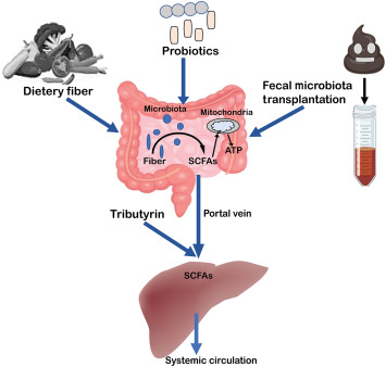
Another strategy is the use of prebiotics (Figure 4). A prebiotic is a “substrate selectively utilized by host microorganisms conferring a health benefit.” Intestinal fermentation of fiber consumed in the diet can also incresase SCFA levels. These dietary carbohydrates escape digestion in the upper digestive tract and are used anaerobically by bacteria of the intestinal microbiota, such as Lactobacillus, Bifidobacterium, and Bacteroidetes. However, other genera, such as Clostridium, can also be stimulated, being particularly important because it is considered a butyrate-producing bacteria. The most common prebiotics are inulin, gluco-oligosaccharides, fructo-oligosaccharides, and β-glucans. Although gastrointestinal microbiota can be affected by fiber and prebiotic intake, individual responses may vary.

Figure 4. The gut microbiota may increase short-chain fatty acid (SCFAs) production as a by-product of the fermentation of dietary fibers, probiotics, or fecal microbiota transplantation. SCFAs are digested and transformed into ATP at the intracellular level, which the cells use as an energy source. The portal circulation receives unmetabolized SCFAs that have crossed the basolateral membrane. SCFAs are used in the liver’s production of ATP and glucose. Just a small proportion of SCFAs that are not used by the liver make it to systemic circulation.

The International Scientific Association for Probiotics and Prebiotics has provided a consensus statement on synbiotics—a strategic combination of prebiotics and probiotics that synergistically promotes beneficial effects on the host’s metabolism and enhances communication between bacterial communities. This approach, known as synbiotics, has the potential to significantly enhance host health by fostering a mutually beneficial association between the host and its microbiota.
To assess the efficacy of probiotics and synbiotics in treating nonalcoholic fatty liver disease, a study by Khan et al examined 12 randomized-controlled trials (NAFLD). The findings demonstrated that probiotics and synbiotics significantly enhanced liver function in patients with NAFLD by lowering aminotransaminase levels and proinflammatory indicators and enhancing liver fibrosis scores. Hence, the study implies that probiotics and synbiotics may be effective NAFLD treatments.

Fecal Microbiota Transplantation

Fecal microbiota transplantation (FMT) is a medical procedure in which feces from a healthy donor are transplanted into a sick patient through the gastrointestinal system to repair the gut microbiota. Inflammatory bowel diseases, Clostridium difficile infections, autism, immune checkpoint inhibitor–associated colitis, hepatic encephalopathy (HE), and other liver ailments may be treatable with FMT. FMT may alter the composition of the recipient’s gut microbiota, and it has shown remarkable efficacy in treating nonmalignant diseases, especially in patients with C. difficile infections. FMT has been acknowledged as the standard treatment for recurrent C. difficile infections.
SCFA-producing bacteria and/or SCFAs improves the effectiveness of chemotherapeutic medications and the host’s response to anti–programmed cell death protein 1 (anti–PD-1) therapy, promoting its therapeutic response and lowering adverse effects. FMT may help restore or improve the number of beneficial bacteria in the gut microbiota of patients with colon cancer, such as Clostridium butyricum, Bifidobacterium, Lactobacillus, and Streptococcus thermophilus, while lowering the number of harmful bacteria.,
HE is a potentially fatal consequence of liver cirrhosis that may cause brain damage and death. Few research studies have investigated FMT in HE, although the outcomes have been promising. FMT increases duodenal mucosal diversity, decreases inflammatory indicators, lowers arterial ammonia concentration, and improves neurologic symptoms. In addition, preclinical research indicates that FMT may prevent HE and enhance liver and intestinal function. FMT may be a potential new therapy for individuals with HE, improving their survival and lowering hospitalizations.
A study examining the safety of FMT to restore gut microbial activity in liver cirrhosis was conducted. Before and after a single FMT, the microbial composition and functional markers of 10 patients allocated to FMT and standard-of-care groups were examined. FMT induced the release of SCFAs and modifies bile acid profiles, restoring metabolic function in the short-term treatments.
In addition, the transplantation of fecal microbiota has emerged as a viable therapy for primary sclerosing cholangitis, and alcohol-induced liver injury

Drugs

Tributyrin, a prodrug of butyrate, enhances butyrate levels in animals. The triglyceride tributyrin comprises three butyrate molecules esterified to a glycerol backbone. On consumption, tributyrin is digested in the gut by pancreatic lipases and esterases to produce butyrate, which may be absorbed and used by the body. Animal studies indicate that tributyrin can increase butyrate levels and improve health outcomes. Egorin et al showed that tributyrin orally administrated to rats and mice induced a peak in butyrate concentration 15 to 60 minutes after administration and remained elevated for approximately 1 hour. Moreover, when butyrate levels in the liver tissue of mice were investigated, it was observed that its levels increased approximately fivefold in the liver of the tributyrin-treated groups compared with the control group. A phase 1 clinical trial was conducted in humans. However, because of its short half-life, the butyrate levels achieved in plasma (0.5 mmol/L) were insufficient to produce a visible effect when administered orally only once daily. When administered three times daily, butyrate levels reached 52 μmol/L, a concentration that induces in vitro effects, but there was great intrapatient variability. Another research study showed that tributyrin/butyrate might inhibit HDAC1, restore carnitine palmitoyl transferase 1A (CPT-1A) expression, and reduce ethanol-induced hepatic steatosis and injury. Similarly, prolonged binge feeding with ethanol has been shown to reduce intestinal tight junction protein level, whereas cotreatment with tributyrin mitigates these effects. Cotreated mice with tributyrin responded less to ethanol-induced TLR mRNA and TNF protein expression in the liver. Another study indicates that dietary supplementation with monobutyrate and tributyrate may be used to alleviate the adverse effects of a high-fat diet. The research discovered that monobutyrate and tributyrate affected the gut microbiome, lowered liver lipids, and decreased succinic acid levels. Similarly, the lipopolysaccharide-induced inhibition of peroxisome proliferator-activated receptor fatty acid oxidation-associated enzymes, fatty acid transport protein, fatty acid binding protein, fatty acid synthesis-associated enzyme, and sterol regulatory element binding protein-1c was inhibited by tributyrin. Tributyrin inhibited the increase in plasma triglyceride, total cholesterol, and low-density lipoprotein cholesterol levels and increased the portal vein butyrate level in mice., Taken together, these studies indicate that tributyrin/butyrate may have therapeutic potential for treating alcoholic and nonalcoholic liver disease.

Argumentative Effects of SCFAs and Butyrate in Liver Diseases

Despite many beneficial effects of dietary fibers and SCFAs from gut microbiota, recent data published on SCFAs indicate that fermentable soluble dietary fibers can also be a potential risk of metabolic disorders in humans., Singh et al identified that enrichment of mouse diet with various fermentable fibers (eg, inulin and pectin) improves conditions of metabolic disorders but induces cholestatic liver diseases and, following HCC, is dependent on the gut microbiota. Occurrences of HCC were correlated with the quantity, type, and length of exposure to soluble dietary fibers consumed in both an inflammatory TLR5 knockout or exposed to high-lipid mouse models. Similarly, SCFAs produced from dietary fiber fermentation exacerbate the inflammation in an IBD mouse model via modulating IL-1β and TLR5 signaling.
When considering the use of butyrate as a treatment for liver disorders, it is important to consider its diverse functions. Despite its potential anti-inflammatory and epigenetic regulatory benefits, butyrate serves as a source of energy for colonocytes and can impact oxidative stress and lipid metabolism. These may have both positive and negative implications for liver disorders., To maximize the therapeutic potential of butyrate in treating liver disorders, further research is needed to understand the underlying mechanisms, determine optimal dosage, and develop targeted delivery strategies.

Conclusion

In summary, this review highlights the numerous beneficial effects of the short-chain fatty acid butyrate on human health. It emphasizes its role in regulating intestinal metabolism, regulating transepithelial fluid transport, inhibiting inflammation, and inducing the epithelial defensive system barrier. The liver receives a significant amount of SCFAs, including butyrate, from the intestines via the portal vein, and recent studies suggest that butyrate has the potential to prevent various liver diseases, such as NAFLD, NASH, inflammation, malignant neoplasms, and liver trauma. Butyrate also shows promise in improving metabolic diseases, like insulin resistance and obesity, while preventing fatty liver disease. Its diverse mechanisms of action, including the inhibition of histone deacetylases and modulation of cellular metabolism, contribute to its multifaceted therapeutic benefits. However, it is important to consider both the beneficial and possible adverse effects of butyrate administration. The future holds promising clinical potential for butyrate in treating liver diseases, and further research is needed to uncover novel intervention targets and methods. Determining the effective doses, timing, and modes of delivery of butyrate administration will be crucial to optimize its efficacy and minimize potential adverse effects. An in-depth understanding of butyrate’s therapeutic properties opens up possibilities for its application in medical care. Future research should focus on identifying specific molecular pathways and targets through which butyrate exerts its beneficial effects. Furthermore, combining butyrate with other therapeutic agents, such as targeted medications or microbiota-based interventions, may lead to synergistic effects and improved treatment outcomes.

References