Neuroendocrinology of mast cells: Challenges and controversies
REVIEW ARTICLE Theoharis C. Theoharides First published: 17 January 2017
https://doi.org/10.1111/exd.13288Digital Object Identifier (DOI)
Abstract
Mast cells (MC) are hemotopoietically derived tissue immune cells that are ubiquitous in the body, including neuroendocrine organs such as the hypothalamus, pineal, pituitary, ovaries, pancreas and uterus where their action is not well understood. Mast cells have historically been associated with allergies because of their rich content of histamine and tryptase, but more recently with regulation of immunity and inflammation due to their synthesis and release of numerous cytokines and chemokines. Mast cells are located perivascularly and express numerous receptors for diverse ligands such as allergens, pathogens, neurotransmitters, neuropeptides and hormones including acetylcholine, calcitonin gene-related peptide (CGRP), corticosteroids, corticotropin-releasing hormone (CRH), β-endorphin, epinephrine, 17β-oestradiol, gonadotrophins, hemokinin-A (HKA), leptin, melatonin, neurotensin (NT), parathyroid hormone (PTH), substance P (SP) and vasoactive intestinal peptide (VIP). Moreover, MC can synthesize and release most of their neurohormonal triggers, including adrenocorticotropin hormone (ACTH), CRH, endorphins, HKA, leptin, melatonin, NT, SP and VIP. Animal experiments have shown that diencephalic MC increase in number during courting in doves, while stimulation of brain and nasal MC leads to activation of the hypothalamic-pituitary-adrenal (HPA) axis. Recent evidence indicates that MC reactivity exhibits diurnal variations, and it is interesting that melatonin appears to regulate MC secretion. However, the way MC change their phenotype or secrete specific molecules selectively at different pathophysiological settings still remains unknown. Mast cells developed over 500 million years ago and may have served as the original prototype neuroimmunoendocrine cell and then evolved into a master regulator of such interactions, especially as most of the known diseases involve neuroinflammation that worsens with stress.
1 Introduction
There are very few studies on the neuroendocrinology of mast cells (MC), especially in the skin.1, 2 A neuroimmunoendocrine circuitry of the “brain-skin connection” 3 has been invoked in many diseases, especially inflammation,[s1] because MC can be stimulated by many neuroimmunoendocrine triggers (Figure 1) and can contribute to a local “hypothalamic-pituitary-adrenal axis.”4 Mast cells derive from haematopoietic precursors5, 6 and mature in tissues in response to stem cell factor (SCF), the ligand of the CD117 (KIT) tyrosine kinase receptor, and local microenvironmental factors7 such as IL-48 and nerve growth factor (NGF).9 The phenotype of the MC can vary considerably with respect to both their content and its responsiveness to stimulatory or inhibitory signals.10
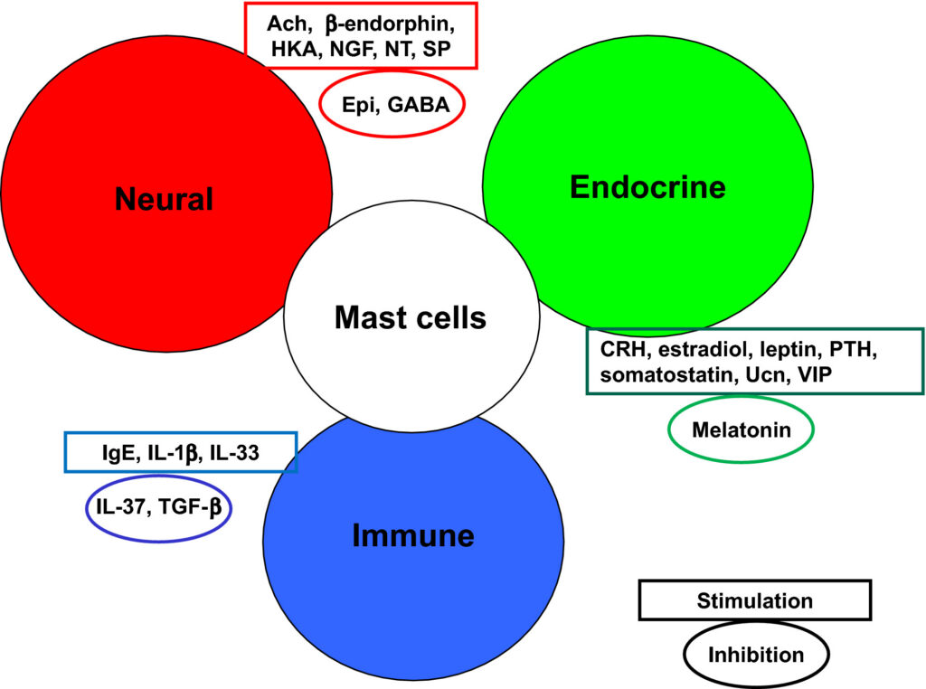
Figure 1 Diagrammatic representation of the stimulatory and inhibitory input on mast cells from the neural, endocrine and immune systems
Each MC contains as many as 500-1000 secretory granules storing numerous biologically active molecules.11 Mast cells are located perivascularly and are typically activated through exposure to allergens that cross-link allergen-specific immunoglobulin E (IgE) bound almost exclusively to high-affinity Fc epsilon receptor 1 (FcεRI).[s2-s5] IgE may have additional regulatory functions on immunity,12 but also on non-immune cells as there is evidence of expression of FcεRI receptors on neurons.13 Mast cells also express receptors for additional diverse ligands,14 including Toll-like receptors (TLR) that can be activated by bacterial and viral products,15 as well as by fungi16 and a variety of toxins.17 Skin MC can also be stimulated by physical stimuli such as pressure, temperature changes and vibration,18 but this mechanism is presently unknown.
Mast cells can also be stimulated by neurotransmitters, neuropeptides and hormones including acetylcholine, calcitonin gene-related peptide (CGRP), corticosteroids, corticotropin-releasing hormone (CRH), β-endorphin, epinephrine, 17β-oestradiol, gonadotrophins, hemokinin-A (HKA), leptin, melatonin, nerve growth factor (NGF), neurotensin (NT), parathyroid hormone (PTH), substance P (SP) and vasoactive intestinal peptide (VIP) (Table 1). Moreover, MC can synthesize and release most of their neurohormonal triggers, including adrenocorticotropin hormone (ACTH), CRH, endorphins, HKA, leptin, melatonin, NT, SP and VIP as discussed later. Mast cell stimulation can be affected by cytokines. For instance, IL-1β triggered selective release of IL-6,19 while IL-33 enhanced allergic MC stimulation;20 moreover, IL-33 had synergistic action with SP inducing vascular endothelial growth factor (VEGF) release21 and acted as “sensors of cell injury,”22 and IL-33 has been considered an “alarmin,”22 acting through MC to alert the innate immune system.23 Other “danger signals” may also stimulate MC.24 However, MC activation ultimately results from an interplay between positive and negative signalling pathways,25, 26 even though there are few if any innate negative regulators of MC known. Mast cell-derived chondroitin sulphate27 and heparin28 have been reported to inhibit MC. Recently, spermine was identified in MC secretory granules29 and we had reported that it inhibits rat MC secretion.30
Table 1. Mast cell neurohormonal triggers
| Trigger | Effect |
| Circadian hormones | |
| Melatonin | ↓ |
| Growth factors | |
| BDNF | ↑ |
| Insulin-like growth factor-1 | ↑ |
| Neurotrophin-3 | ↑ |
| NGF | ↑ |
| PDGF | ↑ |
| SCF | ↑ |
| Metabolic hormones | |
| Leptin | ↑ |
| Neuropeptides | |
| Adrenomedullin | ↑ |
| CGRP | ↑ |
| Hemokinin-A | ↑ |
| Neurotensin | ↑ |
| PACAP | ↑ |
| PTH | ↑ |
| Somatostatin | ↓ |
| SP | ↑ |
| Neurotransmitters | |
| Acetylcholine | ↑ |
| Dopamine | ? |
| Epinephrine | ↓ |
| GABA | ↓ |
| Histamine | ↓ |
| Norepinephrine | ↓ |
| Serotonin | ? |
| Pheromones | |
| Oxytocin | ? |
| Pain peptides | |
| Enkephalins | ↑ |
| β-Endorphin | ↑ |
| Sex hormones | |
| Oestrogen | ↑ |
| Gonadotrophins | ↑ |
| Progesterone | ↓ |
| Testosterone | ↓ |
| Stress hormones | |
| ACTH | ? |
| CRH | ↑ |
| Cortisone | ↓ |
| Urocortin | ↑ |
| Vascular peptides | |
| Endothelin | ↑ |
| VEGF | ? |
| VIP | ↑ |
- ACTH, adrenocorticotropic hormone; BDNF, brain-derived neurotrophic factor; CGRP, calcitonin gene-related peptide; CRH, corticotropin-releasing hormone; GABA, γ-aminobutyric acid; NGF, nerve growth factor; PACAP, pituitary adenylate cyclase-activating polypeptide; PDGF, platelet-derived growth factor; PTH, parathyroid hormone; SP, substance P; VIP, vasoactive intestinal peptide; VEGF, vascular endothelial growth factor.
MC stimulation leads to secretion of numerous vasoactive, neurosensitizing and pro-inflammatory mediators that include preformed, granule-stored mediators such as heparin, histamine, tryptase and TNF secreted immediately through degranulation. Mast cells also secrete lipid-derived mediators synthesized rapidly from membrane-derived arachidonic acid such as prostaglandin D2 (PGD2),31 leukotriene C4 (LTC4)[s6,s7] and platelet-activating factor (PAF).32 MC also produce newly synthesized cytokines such as numerous interleukins (IL), such as IL-5, IL-6, IL-31 and IL-33, and tumor necrosis factor (TNF), and chemokines such as CCL5 and CXCL8, as well as vascular endothelial growth factor (VEGF) released 6-12 hours after stimulation.33, 34 The exact mode of secretion of most of these mediators is still little understood. Triggering through cross-linking of the FcεRI leads to compound exocytosis apparently over 60 minutes, while SP results in more rapid secretion (5-10 minutes) of the content of individual granules.35 However, this picture may be oversimplified. For instance, we showed that MC can secrete the content of individual granules36 or specific mediators, such as serotonin,37 selectively without degranulation.38 We later showed that IL-1 can stimulate selective release of IL-619 as does SCF.39 We also showed that corticotropin-releasing hormone (CRH) can stimulate selective release of VEGF40 as was also reported for PGE2α.41
MC have recently been shown to also have additional means of more rapid and specific communication with other cell types: (i) transgranulation,42 (ii) “polarized” exocytosis of proteolytic enzymes at surface sites called “antibody-dependent degranulation synapse;”43 (iii) formation of nanotubules44, 45 and (iv) secretion of nanovesicles (exosomes)46 that contain many different biologically active molecules,47 in a manner that may be guided by antigens embedded in their phospholipid envelope.48 Such exosomes[s8] could participate in immune49 and neuropsychiatric diseases.50, 51
Consequently, counting the number of intact and degranulated MC, as it typically reported in disease, may be a gross misrepresentation of their potential involvement. Instead, it would be more relevant to investigate the extent of MC mediator release even though MC may otherwise look intact in numbers and degree of degranulation.52, 53
2 Hypothalamic Hormones
Stress contributes to pathological outcomes in various tissues, including skin diseases.54–61,[s9-s11] In fact, stress can worsen allergies,[s12] asthma,[s13] cardiovascular disease[s14] and multiple sclerosis.[s15] Emotional stress is the most common trigger of symptoms in patients with systemic mastocytosis, characterized by increased number and degree of activation of MC.62 Severe stress worsened urticaria pigmentosa and led to increased number of skin MC expressing CRH receptor-1 (CRHR-1).63 The possible role of CRH in cutaneous inflammatory diseases has also been reviewed.64, 65 Psychological stress worsened symptoms in a patient with systemic mastocytosis, who had increased serum CRH levels and whose bone marrow MC also expressed CRHR-1.66 We also reported a case of stress-induced anaphylaxis.67 It is important to note that patients with mastocytosis experience neuropsychiatric problems,[s16-s18] which patients have termed “mastopsychosis.”
Symptoms of both psoriasis (Ps) and atopic dermatitis (AD) worsen with stress.68, 69 These chronic inflammatory skin disorders64, 70, 71 are characterized by severe pruritus,[s19-s20] implicating MC in both Ps72–74 and AD.75–77 It is interesting that maternal psychological problems increase the risk of childhood AD.78 A number of reports indicate more frequent allergies in children with autism spectrum disorder (ASD)79, 80 with food allergies being the most prevalent complaint, often in the absence of elevated serum IgE or positive skin tests.[s21-s23] Large epidemiological studies reported that eczema was strongly associated with ASD and attention-deficit/hyperactivity disorder (ADHD)81 and atopy was associated with risk of both ASD and ADHD.82 Moreover, a large case-control study reported that allergies, asthma and autoimmune disorders, especially Ps, were diagnosed more frequently in patients with ASD than controls.83 An experimental study actually reported that a mouse model of food allergy was characterized by autistic-like behaviour.84
There is strong evidence of the existence of a cutaneous neuroendocrine system,4 involved in skin pathology[s24] through interactions among biogenic amines, catecholamines, melatonin, proopiomelanocortin, adrenocorticotropin hormone (ACTH), β-endorphin and melanin-stimulating hormone (MSH), as well as CRH and related peptides.57, 85 The role of CRH in the skin has been increasingly discussed.64, 65, 86 CRH is typically secreted from the hypothalamus under stress and activates the hypothalamic-pituitary-adrenal (HPA) axis.87 CRH [s25-s27] and CRHR88 gene expression has been documented in rodent and human skin89 and has been shown to contribute to a local HPA axis.90–92 CRH can be released outside the brain from nerve endings,87, 88 but it is also synthesized by skin cells,93 immune cells94 and MC.95 Amazingly, even corticosterone has been localized inside MC secretory granules.96.
MC are juxtaposed to CRH-positive nerve endings in the median eminence,97 as well as during hair follicle formation.98 There is hair follicle-dependent expression of CRH and CRHR in murine skin.99 In fact, CRH can promote MC maturation from local hair follicle-associated progenitor cells.100, 101
Acute stress induces local release of CRH in the skin and increases skin vascular permeability102 through MC103 activation by CRH.104 CRHR-1 gene is expressed in a subpopulation of skin MC,99 and human MC express mRNA and protein for CRHR-1.40, 105 We further showed that skin MC can express CRHR-1, as well as human cultured MC, activation of which induced production of VEGF.40 Stress has also been reported to induce selective release of VEGF,106 an effect that is absent in MC-deficient mice.107 Acute stress and locally secreted CRH103 activate MC68, 103 leading to neurogenic inflammation with subsequent chronic nerve sensitization108 in a “brain-skin” connection.3
In fact, MC have also been implicated in the regulation of HPA axis both in the skin[s28],109 and in the brain.86, 110–112 In particular, histamine was shown to activate the HPA,113 as did MC-derived IL-6114 and CRH.95 Interestingly, unlike its inhibitory effect on other secretory cells, somatostatin triggers MC secretion.115, 116
Mast cells immunoreactive for gonadotrophin-releasing hormone (GRH) increased during courting in the habenula of doves (equivalent to the median eminence in humans).117, 118 MC are absent from the thalamus during pro-oestrus, but are present in the hypothalamus only during the oestrous phase.119 MC in ovarian, uterine and brain tissues change their histamine content throughout the rat oestrous cycle; moreover, brain MC appear to respond to chemosensory cues and are activated during oestrous induction.120
3 Pituitary Hormones
Oxytocin receptors are expressed by MC in the uterus where their activation prevented serotonin uptake.121 Mast cells are also activated by pituitary adenylate cyclase-activating peptide (PACAP), a response augmented by SCF,122 which is an important skin vascular regulator.123
Mast cells are found in the pituitary124 and thyroid125 glands and can regulate thyroid function. Moreover, the incidence of thyroid disease is higher in patients with chronic urticaria.126
It is interesting that PTH is a potent trigger of MC.127
4 Diurnal Peptides
Mast cell numbers and reactivity have been reported to undergo daily rhythmic variations in the rat thyroid gland.128 Moreover, the reactivity of individual MC was further shown to follow a “circadian clock.”129 In this context, it is of interest that MC are present within the pineal gland and melatonin appears to inhibit MC proliferation.130, 131
5 Neurotransmitters
Mast cells have the ability to take up, store and secrete all known neurotransmitters, especially dopamine132, 133 and serotonin.134, 135 Acetylcholine (Ach) at 10−12 M was reported to stimulate skin MC.136
Epinephrine can inhibit TNF release from MC.137 Beta-receptor agonists were shown to be better inhibitors of PGD2 release than cromolyn.138 Such beta-agonists also inhibited IgE-induced histamine release from human gastric MC.139 However, such drugs do not appear to inhibit chronic inflammation in asthma.140
6 Other Neuropeptides
A new human G-protein-coupled receptor MRGPRX2 was recently shown to be activated by cationic drugs and peptides, including SP at high concentrations.141 Nevertheless, SP21, 142–144 and other neuropeptides34 such as NT105, 145–147 and NGF148, 149 also stimulate MC apparently also through specific receptors. In particular, SP can have pro-inflammatory actions acting through its specific NK-1.[s29],150
Psoriasis is also associated with increased skin level of SP, TNF and VEGF. An increased number of contacts between SP-positive nerves and MC have been observed in Ps.55 Moreover, Ps patients with pruritus had increased SP-containing nerve fibre areas and increased number of perivascular degranulated MC compared to Ps patients without pruritus.151 Increased nerve-MC contacts were also reported in the skin of AD patients.152 NT, a brain peptide involved in inflammation,153 stimulates rodent MC through NTR154, 155 to secrete histamine and elevates histamine plasma levels.156 NT administration increases vascular permeability in isolated rat skin157 and in skin blisters in a MC-dependent manner.158 NT is increased in the skin following acute stress, stimulates skin MC and increases vascular permeability.159 It is interesting that NT,160 HKA161 and NGF9 can be synthesized and released from MC.91 NT increases expression of CRHR-1 receptor,162 activation of which by CRH increases allergic stimulation of human MC.163, 164
It should be stressed, however, that there is great tissue variability in MC165 with papers reporting that human skin MC apparently do not degranulate in response to neuropeptides,166 even though their secretion is augmented by specific cytokines such as IL-4 and IL-33.
7 Sex Hormones
Oestrogens and oestrogen receptors[s30] have been implicated in skin pathophysiology as skin disorders are known to worsen during the menstrual cycle.[s31] Human MC express oestrogen receptors167 activation of which increase MC stimulation.67, 168 Oestrogen receptors are also expressed on MC found in bladder169–171 and in lung.172, 173 Oestrogen may activate MC via a non-genomic oestrogen receptor.167 Oestradiol also induced MC migration into the uterus and their degranulation.174 Treatment of mice with luteinizing hormone (LH), follicle-stimulating hormone (FSH) or oestradiol increased the number and extent of MC degranulation in the ovaries.175 Human MC also express progesterone 176, 177 and testosterone178 receptors, but their activation appears to have an inhibitory effect.
Mast cells are present in the human penis,179, 180, prostate181–183 uterus, 184–187, vagina184 and placenta.188 Uterine MC increase during pregnancy and may be important for reproductive processes.[s32] IgE-independent MC activation has been reported to augment contractility of guinea pig,189 mouse190 and human191 myometrium. Activation of MC also leads to angiogenesis in the rat uterine cervix during pregnancy.192
The decidua of women with high levels of stress[s33] and aborted decidua193 contained high number of tryptase-positive MC. We reported high levels of CRH and tryptase in products of conception from women with habitual spontaneous abortions.194 Endometriosis tissue has also been associated with high number of activated MC.195–197 Mast cells were also increased in response to stress that exacerbated endometriosis in a rat model.198 In fact, targeting MC has been proposed as way to treat endometriosis.199 Mast cells have also been implicated in fibrosis, whether it is present in endometriosis or in Hodgkin’s lymphoma.200
8 Metabolic Hormones
Mast cells have been implicated in the metabolic syndrome[s34] and in atherosclerosis.201, 202 Obesity is considered an inflammatory state203 and been associated activation of MC.202,[s35] Moreover, MC secrete leptin,204 which is increased in obesity,[s36] which is increasingly considered an inflammatory disease. Leptin deficiency switches MC to an anti-inflammatory phenotype.205 Insulin and insulin-like growth factor-1 promote MC survival,206 and MC stabilization has been proposed as a means to treat obesity and diabetes.[s37] Skin MC were recently shown to be degranulated in type I diabetes, but to be necessary for healing of diabetic wound ulcers.207
9 Conclusion
MC have been retained throughout the phylogenetic tree208 and may be necessary for survival of the species[s38] by (a) protecting the organism against external triggers;54 (b) fighting pathogens;15, 16 (c) inactivating venoms;17 and (d) regulating immunity,209–211 (e) inflammation,212 (f) the HPA axis110–112, 213, 214 and (g) the gut-brain axis.215, 216,[s39] Some of the key “take-home” messages are listed in Table 2.
Table 2. Main messages
| MC may have functioned as a primitive neuroimmunoendocrine organ |
| MC are found in all tissues, including the brain and endocrine glands |
| MC can synthesize, take up, store and secrete most hormones and neurotransmitters |
| MC are affected by most known hormones and neurotransmitters |
| MC secrete proteases that can either activate pro-peptides or degrade them |
| MC can release mediators selectively through different secretory pathways |
| MC mediators can have autocrine and paracrine effects |
| MC regulate allergic, autoimmune, immune and inflammatory processes |
| MC participate in many diseases affected by stress |
The potential role of MC in immunity217 or autoimmunity218 has occasionally been challenged219 based mostly on the use of certain MC-deficient mice220 that do not necessarily reflect especially inflammatory human conditions.221 It is therefore of critical importance to establish conditions where primary human MC may be cultured together with other cell types, such as human skin fibroblasts,222 to study the development of specific phenotypes.223
It is reasonable to wonder what is the physiological function of MC.[s40,s41] For instance, even though MC bind IgE almost exclusively, it was not known how they do so as IgE circulates and MC develop expression of their FcεRI receptors in the tissues; similarly, MC is the exclusive source of heparin, but MC do not circulate to make heparin available for blood thinning. It was recently shown that perivascular MC extend cell processes through endothelial cell gaps and “capture” circulating IgE;224 this mechanism may also allow them to secrete heparin in the systemic circulation. In this context, it is relevant that endothelin has been shown to stimulate MC, which subsequently degrade it.[S42]
The ability of MC to respond to so many diverse triggers, especially neurohormonal triggers (Table 1), and secrete numerous biologically powerful mediators by utilizing different means of secretion[s43] permits them to participate in many pathophysiological processes such as allergic diseases,[s44-s46] especially in the skin,[s47] but also in the lungs,[s48] innate immunity,7,[s49-s52] autoimmunity[s53,s54] and
inflammation.34, 68, 151, 214, 225,[s55] A case in point is mast cell disorders, especially mastocytosis where patients experience multiple symptoms stemming from the effect of numerous MC-derived mediators affecting many organs.34
10 Remaining Issues
Many questions remain unanswered, especially the way MC change their phenotype or secrete specific molecules selectively at different pathophysiological settings. Other questions include (a) the specific content of human MC secretory granules, (b) the regulation of selective MC mediator secretion, (c) the identification of innate MC inhibitory molecules, (d) the role of MC within endocrine glands, (e) the pathophysiology of uterine MC, (f) the ability of MC to sense odours through the expression of olfactory receptors and (g) the ability of MC to acquire a different phenotype. The latter possibility may permit the use of autologous blood-derived MC progenitors or basophils, or fat or skin-derived MC, to be “transplanted” in the pancreas or substantial nigra for the treatment of type I diabetes or Parkinson’s disease, respectively. Mast cells developed over 500 million years ago and may have served as the original prototype neuroimmunoendocrine cell and then evolved into a master regulator of such interactions, especially as most of the known diseases involve neuroinflammation that worsens with stress.